Introduction
The sternocleidomastoid muscle (SCM) is present in the cervical region and serves as an important landmark in its subdivision into anterior and posterior triangles. As its name suggests, it originates from the sternum and clavicle, and is inserted into the mastoid process. These two heads of origin are replaced by a triangular interval which leads to a surface depression termed the lesser supraclavicular fossa [1].
The present study reports double bellies of the clavicular portion of the sternocleidomastoid; they may reduce this space, leading to overcrowding of the neurovascular structures. This clavicular region has been reported to divide into several parts. In addition, in lower mammals the cleidomastoid section of the sternocleidomastoid is separate from the sternomastoid. Such a variation may be detected in humans as well [2].
The sternocleidomastoid has been utilized as a myocutaneous flap in the correction of oral cavity and facial defects [3], such that aberrations in its structure may be of relevance to medical practitioners in multiple fields.
We aim simply to caution and create awareness among radiologists and surgeons regarding the muscular variants possible for the sternocleidomastoid, a key muscle of the cervical region.
Case Report
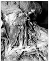
An unusual clavicular head of the SCM was detected during a gross anatomy practical demonstration. The anomaly was unilateral and was discovered on the left side of the cervical region of an adult male cadaver (Fig. 1). The sternal head of the SCM originated as usual from the manubrium sternum; it measured 19.3 cm in length up to its insertion into the mastoid process. The clavicular head of the SCM muscle displayed two bellies, medial and lateral. Both bellies originated from the superior surface of the clavicle, and were separated by a gap measuring 1.6 cm. The width of both bellies was the same, i.e., 1.3 cm. The distance of the medial and lateral bellies at their origin from the sternoclavicular articulation was 3.2 cm and 4.8 cm, respectively. The medial belly of the clavicular head of the SCM fused with the sternal head 10.2 cm from the clavicle. Cranial to the site of fusion, the length of the muscle was 5.4 cm. The lateral belly of the clavicular head fused with the medial belly 8.8 cm from the clavicle; cranial to the point of fusion, the muscle measured 6.2 cm in length. Innervation to both clavicular and sternal heads of the SCM was derived from the spinal accessory nerve. No other neurovascular variations were observed and the other musculature displayed normal morphology.
Discussion
The SCM muscle displays numerous variations, especially in its point of origin [1, 4]. In contrast, however, variations pertaining to insertion are uncommon. An additional sternal head of the SCM has previously been reported [4], while additional clavicular heads were recorded in a separate case [1]. The breadth of the clavicular head of the SCM has been reported previously as narrow or broad. It has been stated that when its origin from the clavicle is broad, it may be subdivided into more than one slip, each separated by a narrow gap [1]. The double clavicular bellies found in the present case report are in accordance with the above statement. Familiarity with these portions of the SCM is vital when one plans to harvest these muscle flaps for reconstructions, such as during performance of parotid surgery, where this muscle flap may prove vital in covering surgical defects and will likely avert Frey's syndrome [1]. Surgeons must therefore remain vigilant of such muscular variations in planning head and neck surgeries.
An embryological basis for the supernumerary clavicular head may be the separation or division of mesoderm at the sixth brachial arch.
Whether these gross anatomical findings can be corroborated with clinical conditions such as wry neck is a topic of speculation and has not yet been systematically evaluated. Nonetheless, the presence of an additional muscle belly is likely to increase the propensity of the muscle to undergo spasm, either congenitally or acquired.
An earlier finding reported by Bergman et al. [2] stated that the SCM is disposed in two layers, superficial and profound, that subdivide into five parts. The superficial portion of the SCM may have sternomastoid, sterno-occipital, and cleido-occipital subregions. Additionally, the profound part may display sternomastoid and cleidoomastoid subregions.
Another previous description identified superficial and deep layers, where the superficial portion exhibited sternomastoid and sternocliedooccipital parts but the deep layer the cleidomastoid part only [5]. An incidence of 33% has been recorded for cleido-occipital muscle [6]. Our report differs from the above with respect to observation of two subportions of the SCM's clavicular head. Moreover, the SCM was not disposed in superficial and deep layers in our case.
The current case description finds similarity with an earlier finding of an additional (third) head originating from the superficial surface of the middle third of the clavicle. However, this head passed deeper than the normal clavicular muscle head to blend with the SCM fascia [7]. In contrast, the lateral belly of the SCM in the cadaveric specimen discussed here was not observed to pass deeper than the normal belly; indeed, all three masses combined together toward the middle of the neck.
Head and neck plastic surgeons will benefit most from a discussion of potential anomalies in the SCM muscle. The SCM has been implicated in various reconstructions of the head and neck region; it may be utilized as a myocutaneous flap in reconstructing the oral floor, as a suture line to protect the carotid arteries, or, along with a portion of the clavicle, to reconstruct the mandible [8]. Furthermore, the utility of this additional clavicular head in parotidectomy has been previously addressed [9].
The strategic position of the additional belly found in the present study, in relation to vital cervical neurovascular structures, must be appreciated by surgeons operating in this region. Therefore, identification of such aberrations prior to contemplating invasive procedures of this region is essential. Another safe option is to reconstruct the SCM intraoperatively to prevent complications.
An interesting case described in a previous work involved an additional sternal and three accessory clavicular heads bilaterally. An accurate and appropriate anatomical knowledge for anaesthetists is mandatory before attempting a central venous catheterisation approach for internal jugular vein (IJV) cannulation. This route is preferred due to its low tendency to cause pnemothorax [10]. Therefore, in such cases considerable narrowing of the normal gap between the two heads of the SCM would jeopardize operative interventions in this area.
We further infer that the presence of an additional clavicular belly narrows the minor supraclavicular fossa of the neck, leading to cumbersome IJV cannulation. This difficulty during cannulation can accidentally puncture the neighbouring neurovascular structures thereby leading to haematoma formation or resulting in neural deficits.
Since the SCM and trapezius originate from the same myotome in the occipital region, just caudal to the last brachial arch, they are often fused, especially at the site of insertion [11, 12]. This myotome segregates into the vertical SCM and dorsal trapezius. Occasionally, therefore, the margins of these two muscles make contact with each other [11, 12]. Moreover, HOX D4 and somitic mesoderm contributes to the development of these muscles where they are connected to skeletal elements only by posteriorotic neural-crest derived connective tissue. Since HOX D4 and somites contribute muscle cells to branchial neck muscles, these myoblasts seem to associate with neural-crest derived muscular connective tissue [13].
The presence of these anomalous variants in relation to SCM muscle may be attributed to phylogenetic processes. The cleidomastoid portion of the SCM is reported to be distinct from the sternomastoid part in animals such as rabbits and crocodiles. Such a situation may be found in humans as well [14]. Therefore, we wish to emphasize that a sound knowledge of comparative anatomy pertaining to lower mammals may prove useful in the comprehension of muscular variants of the cervical region.
The SCM serves as an important soft tissue landmark in the interpretation of magnetic resonance imaging scans. Moreover, the presence of this variation could alter the dosage of botulinum toxin injections administered to patients with irradiation-induced muscle spasm. Patients with supernumerary muscles would require higher doses of such an injection [15].
The SCM provides the mechanical basis in a wide variety of head movements and is considered an accessory muscle in respiration [16]. The presence of two bellies at the clavicular head of the SCM, detected unilaterally in the current study, may possibly influence muscular biomechanics. The authors speculate that the additional clavicular belly as presented herein may provide a functional advantage, as it serves to augment the usually present clavicular head. It is also pertinent to suggest that the relatively superficial location of the additional bellies of muscles such as the SCM enhances its suitability for its use in reconstructive operations.
In view of the current study, it may be stated that awareness and precise knowledge of muscular variants of the SCM is of relevance to surgeons, radiologists, and anaesthetists in their clinical practice.


 PDF
PDF ePub
ePub Citation
 Print
Print


 XML Download