

 PDF
PDF ePub
ePub Citation
 Print
Print


Introduction
The axillary region is a clinically important area because it possesses neurovascular and lymph node structures associated between the neck and the upper limb. Anatomical variations of axillary muscular slips may cause obstructions of axillary vessels and nerves. Muscular variations in the axillary region may be involved in thoracic outlet syndrome, shoulder instability, development of lymph edema of the upper limb, and surgical interventions such as breast surgery [1-3]. By shoulder magnetic resonance imaging examination, Guy et al. [4] suggested that the axillary arch can cause lymph node concealment and brachial plexus impingement.
In general, the muscular variation of the axilla (known as axillary arch) is described as the presence of variant thin muscular/tendinous slip (also called Langer's muscle) arising from the medial border of the latissimus dorsi with no insertion onto typical floor of intertubercular groove of the humerus [5].
The variant origins and insertions of the axillary arch muscles have been described [6-8]. In most cases the origin of a classical variant muscular slip arises from the latissimus dorsi muscle whereas the insertions varied from single to other structures at multiple sites [7, 8]. In the present observation, we report duplicated axillary arch muscular variations that arise from the latissimus dorsi muscle but have two different insertions. This is a type of axillary arch that has not been previously described.
Case Report
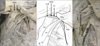
During the routine dissection of embalmed cadavers for teaching medical students at the Medical Gross Anatomy Laboratory in the Department of Anatomy, Faculty of Medicine, Khon Kaen University, the upper limbs were dissected and observed carefully to study the compartments of the pectoral and axillary regions. Unexpectedly, in a 33-year-old male cadaver, we found two variant muscular slips arising from a common inferolateral border of the right side latissimus dorsi muscle (Fig. 1). We considered these muscular variations duplicated axillary arch muscles (also called Langer's muscle). These muscular slips were distinguishable as long and short muscular bands (Fig. 1A, B). The long slip is about 13.5 cm in length, 1.7 cm in width measured at the broadest point, and 0.4 cm in thickness. The short one is approximately about 9.1 cm in length, 1.6 cm in width, and 0.4 cm in thickness. These two slips arise from the same origin (inferolateral border of the latissimus dorsi), but their insertions are totally different (Fig. 1). For the origin location, the short and long muscular slips arise near the location of the second and third ribs, respectively. The short muscular slip inserts into the deep fascia covering on the pectoralis minor, whereas the longer one runs on/into the aponeurosis of pectoralis major (Fig. 1C). We could not find separate innervations and arterial supplies of each muscular slip. However, small branches of the first and second intercostobrachial nerves were present under this muscle arch around the first intercostal space. It seemed that thoracodorsal vessels and nerve, lateral thoracic vessels and long thoracic nerve, and lymph nodes were closely located underneath the muscular slips. In contrast, the anomalous muscles of the left side axillary region were not observed.
Discussion
The axillary region is a complicated area that contains the neurovascular bundles and lymphatic glands, surrounded by a large quantity of fibrous capsules and adipose tissue. Therefore, it is possible that axillary arch muscles might cause lymph node concealment or brachial plexus impingement. In addition, the muscular variations in this region must be considered before performing any surgical intervention such as axillary lymphadenectomy for breast carcinoma [3, 9]
Most muscular variation of the axilla is the presence of unusual muscle slips, typically called "axillary arch muscle." Classically, the origin of variant axillary muscular slips comes from the border of either latissimus dorsi or pectoralis major, whereas the insertions vary as discussed in Lama and Tamang [8]. The latissimus dorsi-arising axillary arch muscle inserts into the fascia covering the common tendon of short head of biceps brachii and coracobrachialis [6], tendon of pectoralis major [10, 11], or aponeurosis of the coracobrachialis muscle [11]. In addition, Lama and Tamang [8] reported a rare axillary arch muscle slip that originated from the latissimus dorsi and inserted onto the medial epicondyle of the humerus and the medial intermuscular septum. Multiple insertions (to pectoralis major, pectoralis minor, and coracoid process) of an axillary arch muscle originating from latissimus dorsi have been reported [7].
We present a rare case found in a Thai cadaver where the two axillary muscle slips (short and long bands) raised from a common inferolateral border of the latissimus dorsi and inserted into two different sites (Fig . 1); the fascia covering on the pectoralis minor (for the short band) and the aponeurosis of pectoralis major (for the long band).
The origins of these anomalous muscular fibers obviously arose from the latissimus dorsi muscle. The morphogenesis of these muscles cannot be exactly explained. It is possible that duplicated axillary arch muscles unusually developed from the latissimus dorsi muscle innervated by thoracodorsal nerves. In the present report, the innervations of duplicated axillary arch muscles by the thoracodorsal nerves were not observed. However, we found that small branches of intercostobrachial nerves seemed to innervate this muscular arch.
Axillary arch muscle is implicated in various clinical complications such as neurovascular compression syndrome [12]. These may include the entrapments of axillary vein, musculocutaneous, median, and ulnar nerves [6, 13]. Furthermore, it is possible that axillary arch causes functional disturbances including shoulder elevation and hyperabduction syndrome because of the restriction of axillary muscular slip.
In conclusion, the unique formation of such an axillary arch with two different insertion sites has never been reported. Although we could not prove any functional disorder because the study was done in an embalmed cadaver, this variation might result in the inability to completely elevate the shoulder. The variations in formation of the axillary arch muscles are very important information for surgeons before performing any serious surgery in the axillary region.
 XML Download