

 PDF
PDF ePub
ePub Citation
 Print
Print


Introduction
The teres major (TerMa) muscle is a scapulohumeral muscle, in which its tendon transfer improved the arm function in patients who suffered from massive rotator cuff tears, by a Salvage procedure [1, 2]. In a novel technique, TerMa and latissimus dorsi (LD) tendon transfer through a deltopectoral exposure, has also been considered in treating complex motor deficiencies around the shoulder [3-5]. TerMa originates from dorsum of the inferior angle (inferolateral third) of scapula and inserts into the medial lip of bicepital groove of the humerus. In common primary actions, these two muscles extend, adduct, and medially rotate the arm.
According to the text in Gray's Anatomy [6], it is stated that the variations of muscular slip fusions between TerMa and LD have been observed; however, the variation of the teres major insertion is very rare. For an effective surgery of the irreparable cuff tears, the knowledge on variations of either TerMa or LD is important to guide the clinicians during surgical procedures. In this report, we present a variation of insertion of the TerMa muscle directly attached at the tendon of LD and attempted to discuss the possible anatomical and clinical significances.
Case Report
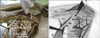
During routine dissection (at the Medical Gross Anatomy Laboratory in the Department of Anatomy, Faculty of Medicine, Khon Kaen University) of the embalmed cadavers for teaching medical and paramedical students, the shoulder and axillary regions were dissected and observed carefully to study the normal origins and insertions of the scapulohumeral muscles. We found an uncommon insertion of the right TerMa with typical origin in the embalmed cadaver of a 33-year-old Thai male (Fig. 1A). As an observation, TerMa muscle fibers inserted directly at the supero-medial border of the latissimus dorsi tendon (LDt) (Fig. 1A, B) and the medial lip of the bicipital groove, where this muscle usually attaches was empty. Compared to the normal TerMa's insertion, it was noted that the terminal tendon of TerMa was not observed. The width at the insertion border of TerMa muscle fibers to supero-medial border of LDt was approximately 7.2 cm (Fig. 1A, black arrows). This variant TerMa was innervated and supplied by lower subscapular nerve and circumflex scapular artery. In contrast, this variation of the left TerMa was not observed.
Discussion
Basically, TerMa's tendon inserts into the medial lip of bicipital groove and LD's tendon attaches at the floor of the bicipital groove of the humerus. Although it has been found that muscle fibers of TerMa and LD could be fused to each other [6], the reports about the variant types of TerMa and LD are limited. Herein, we report that all muscle fibers of TerMa directly fused to the tendon of LD at the supero-medial border (Fig. 1A, B). It is possible that the development of the TerMa and LD muscles was very closely related. In addition, it could have the impairment of TerMa's tendon formation and the errors in the migration of the myoblast, which built up the TerMa possessing of the similarity in the direction, the action, and innervation between the TerMa and LD. Basically, the width of the TerMa tendon is shorter than that of its muscle fibers. In tendon dimensions, it has been reported that the width at insertion of TerMa tendon was 4.0 cm (range, 3.3 to 5.0 cm) [4]. Herein, we report that the width at insertion border was a 7.2 cm (Fig. 1), which was longer than the previous report [4]. It could be explained that the width at insertion border of TerMa in our case was only muscle fibers (note: there was no terminal tendon of TerMa [Fig. 1]).
The TerMa and LD, together, are responsible for the normal actions of adduction and medial rotation of the humerus and assistance in the extension of the arm. The TerMa is also important for stabilization of the humeral head in the glenoid cavity, resulting in steady of the head in its socket. In the present case, the anatomical variation of the TerMa's tendon may cause slightly abnormal primary actions of the arm because no tendon of TerMa inserted on medial lip of bicipital groove of the humerus.
In treating the rotator cuff deficiency, the tendons of LD, teres major, pectoralis major, deltoid, or trapezius muscle are commonly considered for tendon transfer surgeries, including the Salvage procedure [3, 5, 7, 8]. Among the muscles mentioned above, the TerMa, has been demonstrated to have the most functional activation after transfer surgery [1, 2]. Moreover, the use of LD presented in this case for breast reconstruction, cardiomyoplasty and other reconstructive surgery shoud be considerd. As a clinical significance of the TerMa, knowledge on variations of this muscle, especially its tendon insertion, is very important for surgeons, in order to avoid misinterpretation of normal condition. Therefore, a very rare variation of the TerMa insertion in this presentation is necessary to be recognized.
 XML Download