Introduction
Variations in the upper limbs, especially in its nerves, vessels and muscles, are common and have been reported by many authors [1-3]. In this regard, variation in the formation of brachial plexus (BP) during its course, branches and distribution is of great interest for all the clinicians. Knowledge of such anatomical variations of BP and its branches in the upper limb is important because these variants could be injured during surgical procedures, producing unusual clinical symptoms.
The median nerve (MN) is formed by the union of lateral and medial roots from lateral and medial cord of BP respectively. It then courses downwards in front of the arm; crosses the brachial artery from lateral to the medial side and finally enters into forearm by crossing the elbow joint. Usually MN does not branch to the arm muscles except to one of the forearm muscle, pronator teres whose nerve arises from the MN before the MN crosses the elbow [4]. All the anterior arm muscles, coracobrachialis (CB), biceps brachii and brachialis are supplied by musculocutaneous nerve (MCN) branch of lateral cord of BP, and after supplying these muscles, the MCN continues as the lateral cutaneous nerve of forearm (LCNF) to supply the skin of lateral forearm.
The aim of the present study is to report the bilateral absence of MCN and the variant innervation of the front muscles of arm and lateral side of skin of forearm from MN.
Case Report
During routine upper limb dissection for first year medical students, the BP of 43-year-old female cadaver showed the complete absence of MCN and variant branches of MN in the arm of both the upper limbs.
Right side
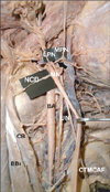
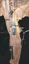
In the right axilla, the lateral cord of BP had three branches i.e. lateral pectoral nerve, nerve to CB and lateral root of MN. The lateral pectoral nerve pierced the clavipectoral fascia and supplied the pectoralis major as usual but during its course it communicated with the medial pectoral nerve (MPN) of medial cord of BP (Fig. 1). The MCN was completely absent. The nerve to the CB was a small motor twig arising directly from the lateral cord which innervated the CB muscle. The lateral and medial root of MN formed the MN proper and descended between the biceps brachii and brachialis muscle. It then crossed the brachial artery from lateral to medial and finally entered the cubital fossa. In the arm, deep in the biceps brachii, the MN gave two branches: a direct branch to the biceps brachii and another long branch which, after supplying the brachialis muscle, continued as the LCNF and supplied the lateral side of the skin of forearm. The medial cord also varied in the origin of medial cutaneous nerve of arm and forearm arising from a single common trunk, which became separated in the middle of the arm (Fig. 2).
Left side
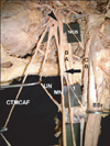
The lateral cord of BP in the left axilla branched four times: the lateral pectoral nerve; nerve to CB and lateral root of MN and an additional lateral root of MN. The MCN was absent and the nerve to CB was a small twig which supplied and terminated in the muscle itself (Fig. 3). No communication between lateral and MPNs was found.
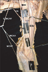
The MN was formed by two lateral roots from the lateral cord and one from medial root from the medial cord. After the formation, the left MN had the same course as the right side. During its course between the biceps brachii and brachialis, the MN split off a single long branch from which branches to the biceps brachii and brachialis were derived. After supplying these two muscles it continued as LCNF, which supplied the lateral side of the skin of forearm. The medial cutaneous nerve of arm and forearm arose from a common trunk, and separated in the middle of the arm, similarly to the right side (Fig. 4).
Discussion
Anatomical variations of the BP have been reported by several authors. The MCN and the MN are the two major nerves which have numerous variations in their formation and branching patterns. The MCN is one of the three terminal branches of lateral cord of the BP which after piercing the CB, innervates the musculatures of anterior compartment of arm and continues as LCNF [4].
Variations of the MN and absence of the MCN have been extensively studied by many authors [1, 5-7]. Variations in the formation, distribution and termination of the MCN have been described by Bergman et al. [8] who reported that 90% of the MCN arises from the lateral cord while in 2% of the cases it may arise from the MN or may be completely absent. Le Minor [6] and Gümusburun and Adigüzel [7] classified five types of variation, with the 5th type being the complete absence of MCN, where anterior arm muscles will be supplied by the MN. Similar cases of the absence of the MCN and the unusual branches of the MN have been reported in literature [3, 6]. Usually if the MCN is absent, the fibers of the MCN are en-routed through the lateral root of MN which takes the role of the MCN in supplying all the anterior arm muscles and lateral side of the skin of forearm [6]. Our present case had complete absence of the MCN coinciding Le Minor's [6] and Gümusburun and Adigüzel's [7] 5th type of classification, but the innervation of the anterior compartment of arm muscles were unique in that none of the muscles were supplied by the MN. Only the branches to biceps brachii and brachialis were derived from the MN through its lateral root and the nerve to the CB arose directly from the lateral cord of the BP. However direct branches to the CB from the lateral cord [9] and the lateral root of the MN [10] are also been reported in addition to the presence of the MCN.
On the left side we found a variation in the formation of the MN with an additional lateral root which ran downwards, medially and joined the lower part of the medial root of the MN. Cases of MN formation with additional roots have been reported by several authors [3, 11, 12]. The additional lateral root with the absence of the MCN has been reported. This unusual root was found in our case, crossing the third part of axillary artery, and it may convey the fibers of the MCN but at the same time there is a risk of compression of the artery, lessening the blood supply to the left upper limb [11].
Anomalous communications in the branches of BP, like communication between medial and lateral pectoral nerve, median and ulnar nerve, medial root of MN and additional lateral root of MN have been reported in the literature [3]. The present case illustrates communication between medial and lateral pectoral nerve on the right upper limb. No communication was found between medial cutaneous nerve of the arm and forearm; instead both the nerves arose from a single common trunk which separated in the middle of the arm.
The present study can be incorporated with recapitulation of evolutionary aspects because in lower vertebrates like amphibians, reptiles and birds there is only one trunk equivalent to the MN in the thoracic limb. The absence of MCN and the innervation of the anterior arm muscles by MN may also be interpreted on the developmental basis of the upper limb muscles. The upper limb muscles in man develop from the mesenchyme of paraxial mesoderm while the axons of spinal nerve supplying it grow distally to reach the muscles and skin of upper limb bud [3, 13]. Any variation in the coordination of muscle development and its neuronal signalling results in the anomalies of the upper limb musculature and its innervation.
In summary, this paper is to report the absence of the MCN, nerve to CB and innervation pattern of MN in the arm might be of significance in diagnostic clinical neurology. Knowledge of such variations of the BP as found in our study may provide additional anatomical information for the clinicians during diagnosis of unusual clinical symptoms and for surgeons to avoid damage to these nerves, during surgical exploration of axilla and arm region, shoulder reconstructive surgeries and anterior surgical approaches of the upper limb.


 PDF
PDF ePub
ePub Citation
 Print
Print


 XML Download