

 PDF
PDF ePub
ePub Citation
 Print
Print


Introduction
Anatomical variations in the upper limb are frequent, but the coexistence of multiple combined neuromuscular and vascular variations are rare. These variations are worthy of note for clinicians since they should be kept in mind in surgical and diagnostic procedures. In the present study we aimed to describe the co-existence of a high originated radial artery, supernumerary heads of the coracobrachialis and biceps brachii muscles and communication between the median and musculocutaneous nerves. The presence of this co-existence was discussed from embryological and clinical points of view.
Case Report
Methods
During a routine cadaveric dissection at the Department of Anatomy, Akdeniz University Faculty of Medicine, Antalya, Turkey, we found multiple variations on the left side of a 51-year-old Turkish male cadaver. The arm was dissected carefully to display all structures. All other related structures were also exposed.
Results
Arterial variations
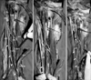
The brachial artery was split into two main branches at the level of middle third of the humerus. The radial artery coursed downwards and crossed the median nerve from medial to lateral side (Fig. 1A). Branches and terminations of the radial artery were normal in every aspect. The remaining branch coursed downwards as the ulnar artery. It passed under the bicipital aponeurosis and then gave its main branches.
Nervous variations
A communicating branch originated from the median nerve at the level of upper third of the humerus, coursed downwards through two supernumerary heads of the biceps brachii muscle and finally joined to the musculocutaneous nerve at the level of lower third of the humerus (Fig. 1B). The united nerve trunks joined to become the lateral antebrachial cutaneous nerve. There was no variation in the course and distribution of the lateral antebrachial cutaneous nerve (Fig. 1C).
Muscular variations
The reported coracobrachialis muscle consisted of three heads. The first two heads had originated from corocoid process as superficial and deep heads and were inserted in the middle third of the humerus as usual insertion site (Fig. 1A-C).
The musculocutaneous nerve was sandwiched by both of these two heads. The remaining head of the coracobrachialis muscle was a capsular head and had originated from articular capsule of the glenohumeral joint (Fig. 1C).
The biceps brachii muscle had two supernumerary heads. The first supernumerary head had originated from the insertion site of the coracobrachialis muscle as a united tendon (Fig. 1A). This head united to the medial edge of the short head of the biceps brachii muscle. The second supernumerary head had originated from the upper third of the humerus (Fig. 1A). This head was united to the medial edge of the long head of the biceps brachii muscle (Fig. 1C).
Discussion
The biceps brachii muscle presents in a wide range of variations. Supernumerary heads of the biceps brachii muscle have been widely studied regarding their origin, insertion, size, innervation and racial differences [1-5]. The supernumerary heads of the biceps brachii muscle have been described as part of either a three-, four-, five-headed or even seven-headed biceps brachii muscle [4, 6]. The prevalence of a supernumerary head of the biceps brachii muscle varies among populations, such as Chinese (8%), European white (10%), African black (12%), Japanese (18%), South African blacks (21%), South African whites (8%), and Colombians (38%) [1, 3, 4]. The four-headed biceps brachii variation is less frequent with a reported prevalence of 0.18-2.75% [7].
Supernumerary heads of the biceps brachii muscle may arise from the articular capsule of the glenohumeral joint, lesser tubercle, coracoid process, pectoralis minor tendon, humeral shaft, tendon of the pectoralis major muscle, or the crest of the greater tubercle [1, 6, 8-11]. Recently, Rodríguez-Niedenführ et al. [3] studied on a series of 350 arms and classified the supernumerary heads of the biceps brachii into three different types: superior, inferomedial, and inferolateral humeral head. They found five cases (1.5%) with a supernumerary head that originated from the surface of the humerus between the lesser tubercle and the attachments of the coracobrachialis and brachialis muscles and fused with the muscular fibers of the short head of the biceps brachii muscle at its union with the long head.
The presence of the supernumerary heads of the biceps brachii muscle has been associated with variations of the surrounding muscles. El-Naggar and Zahir [12] described that two bellies of the coracobrachialis muscle associated with a third head of the biceps brachii muscle, although the coracobrachialis muscle was found to have a normal origin, and the short head of the biceps brachii muscle had separate bellies. From this point of view, the present case also had multiple variations (arterial, nervous and muscular) of which they were encountered unilaterally.
Anomalies of the coracobrachialis muscle are common [1, 13]. Numerous studies have documented variations of the form and origins of the coracobrachialis muscle. The reported morphological variations of the coracobrachialis muscle that included accessory slips inserting to the medial epicondyle of the humerus, medial supracondylar ridge, medial intermuscular septum, the lesser tubercle and a supernumerary head passing over the shoulder joint [14]. The morphological variations of the coracobrachialis muscle may be due to failure of muscle primordia disappearing during embryological development [15]. The origin of morphologic variations of the coracobrachialis muscle may be explained on the basis of the embryogenesis of the muscles of the arm. The intrinsic muscles of the upper limb differentiate in situ from the limb bud mesenchyme of the lateral plate mesoderm. At a certain stage of development, the muscle primordia within the different layers of the arm fuse to form a single muscle mass; thereafter, some muscle primordia disappear through cell death [15]. Failure of muscle primordia to disappear during embryologic development may account for the presence of the supernumerary heads of coracobrachialis muscle as reported in this case. The coexistence of these variations may be the result of an abnormal embryological formation of the limb muscles, peripheral nerves and arteries.
In most species, the coracobrachialis muscle has three portions: the longus, medius and brevis. In humans, the medius and longus fuse to form the coracobrachialis muscle [1]. The original third head of the muscle, termed coracobrachialis brevis, occurs rather rarely [1, 13]. Kyou-Jouffroy et al. [16] described three portions in coracobrachialis muscle that originated from the coracoid process and inserted into the medial epicondyle of the humerus (coracobrachialis longus or superficialis), humeral diaphysis (coracobrachialis medius) and humeral neck (coracobrachialis profundus or brevis). Mori [17] found that coracobrachialis was completely separated into its superficial and deep layers in eight arms (16%) and incompletely in four arms (8%).
Variations in the connections of musculocutaneous and median nerves have been classified in many ways. In two separate reports Testut [18] and Monden [19] reported that the incidence of the communications between musculocutaneus and median nerves was 36.2% (38 of 105 cases), and 27.5% (33 of 120 cases), respectively. Numerous communications between the median and musculocutaneous nerve have been classified regarding their presence, formations and racial differences [20-22]. In the present case we found that communication originated from the median nerve at the level of upper third of the humerus, coursed downwards through two supernumerary heads of the biceps brachii muscle and finally joined to the musculocutaneous nerve at the level of lower third of the humerus. A clinician's knowledge of musculocutaneous-median nerve communication is important while evaluation of clinical neurophysiology, planning a surgery after trauma and understanding of median and musculocutaneous nerve dysfunction [23-25].
Presence of the multiple variations has also been reported in a previous report [26]. In our previous study we found a high originated radial artery accompanied by muscular and neural abnormalities (three-headed biceps brachii, absence of the palmaris longus muscle, and communication between median and musculocutaneous nerves). In the present case, there was a four-headed biceps brachii muscle and three-headed coracobrachialis muscle accompanied by high originated radial artery and communication between median and musculocutaneous nerves. Rodríguez-Niedenführ et al. [3] reported a case with four-headed biceps brachii muscle of the right arm, but they did not observe any connection between the median and musculocutaneous nerves. Although it is not usually reported, the presence of the multiple variations is a common case that encountered during educational and diagnostic procedures. We think that the presence of the multiple variations is worth of note not only for anatomist but also for clinicians. Because the upper extremity is a frequent site of injury, various surgical and invasive procedures are performed in this region; consequently, it is of utmost importance to be aware of such variations. The anatomical variations and abnormalities of the muscles of the upper limb have become significant because of physicians may encounter such abnormalities during imaging with computed tomography and magnetic resonance. Also, these variations are important in order to define the anatomical features of each in relation to the clinical diagnosis and for surgical procedures [27]. Therefore, it should be kept in mind during routine dissection studies, and surgical/diagnostic procedures.
 XML Download