

 PDF
PDF ePub
ePub Citation
 Print
Print


Introduction
The biceps brachii muscle shows variations in the number of heads, with an estimated 9-22% of all people having a supernumerary head [1]. The most common variation is a third head, but four, five, or even seven heads have been reported [2-4]. The supernumerary heads of the biceps brachii muscle have clinical importance, as they may confuse a surgeon who performs a procedure on the arms or they may cause compression of neurovascular structures in the upper limbs [5]. Although supernumerary heads of the biceps brachii muscle have been frequently reported, bilateral asymmetric occurrence of supernumerary heads is relatively rare. We recently observed an intriguing variation of the biceps brachii muscle during a cadaveric dissection.
Case Report
Methods
During a routine cadaveric dissection at the Department of Anatomy, Chonnam National University Medical School, Gwangju, Korea, we found three accessory heads of the biceps brachii muscle on the right side of the arm and an anomalous third head of the biceps brachii on the left side of the arm of a 50-year-old male Korean. The arm was dissected carefully to display the full length of the biceps muscle from proximal to distal attachment. All other related structures were also exposed. The additional heads were examined for the origin and course at the lower end. Appropriate photographs were taken.
Results
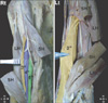
In this case, three accessory heads of the biceps brachii muscle were observed on the right side of the arm. These heads originated from the body of the humerus at the insertion site of the coracobrachialis muscle and inserted into the distal part of the short head of the biceps brachii, proximal to the distal in order. We also observed that the musculocutaneous nerve had a characteristic branching pattern between the third and fourth heads (Fig. 1).
In addition, an anomalous third head of the biceps brachii muscle was observed on the left side of the arm. The third head originated from the insertion site of the coracobrachialis muscle on the humerus and was fused with the common bulk of the muscle, well before the bicipital tendon and its aponeurosis. This additional head was supplied by a branch of the musculocutaneous nerve (Fig. 1). No additional anomalies were found on the remainder of both upper limbs of the same cadaver.
Discussion
The biceps brachii muscle presents a wide range of variations. This anomaly varies among populations, such as Chinese, 8%; European white, 10%; African black, 12%; Japanese, 18%; South African blacks, 21%; South African whites, 8%; and 38% in Colombians [6]. The anomalies can manifest as a cluster of accessory fascicles arising from the coracoid process, pectoralis minor tendon, proximal head of the humerus, or articular capsule of the humerus [7]. The most common variation is the muscle arising from the shaft of the humerus itself, also known as the humeral head or the third head of the biceps brachii muscle [8].
The presence of a supernumerary head of the biceps brachii muscle might increase its kinematics. The biceps is known for its powerful elbow flexion action (secondary to brachialis) when the forearm is supinated. It also acts when rapid supination is required. Anatomists consider that additional biceps heads, as observed in this case, may increase the power of flexion and the supination component of the elbow. However, no attempt to amalgamate the relationship between additional heads and muscle strength has been made.
Embryological observations by Testut described this type of variation with the third head of the biceps brachii as a portion of the brachialis muscle supplied by the musculocutaneous nerve, in which its distal insertion has been translocated from the ulna to the radius [9]. However, in the present case, additional heads of both sides originated from the anteromedial surface of the humerus distal to the insertion site of the coracobrachialis muscle. Therefore considering their origin they may be remnants of the long head of the coracobrachialis muscle, an ancestral hominoid muscle [10].
The existence of accessory muscles in the arm and fore-arm may confuse surgeons during procedures, cause compression of neurovascular structures, or lead to variation of normal mechanical actions. Variations in the heads of the biceps brachii muscle have already been reported to cause compression of surrounding neurovascular structures and lead to erroneous interpretation during routine surgeries. The branching pattern of the musculocutaneous nerve may be clinically important, as the nerve is subjected to compression by the accessory heads. The knowledge of such variations may be important for surgeons operating on the arm and for clinicians diagnosing nerve impairment.
We report a peculiar origin of bilateral asymmetric supernumerary heads of the biceps brachii muscle in the present case. Variations in the heads of the biceps brachii also have clinical importance, as they may confuse a surgeon during surgical procedures.
 XML Download