

 PDF
PDF ePub
ePub Citation
 Print
Print


Introduction
A number of authors have reported the numerous variations of the wrist flexor muscles [1-7]. Flexor digitorum süperficialis (FDS), palmaris longus muscles are a well known example of flexor compartment of the forearm [8-15]. The accessory flexor carpi ulnaris (AFCU) had been previously described by a few authors [16, 17].
These muscles are usually asymptomatic and discovered incidentially during anatomical dissection or at operation [18-20].
However, they may simulate a ganglion [21] or a soft tissue tumour [22] or cause pressure neurits and produce symptoms such as a carpal tunnel syndrome [14, 23].
The FCU, usually arises by two heads, humeral and ulnar, connected by a tendinous arch. The humeral head originate from the medial epicondyle and ulnar head from the medial margin of the olecranon and proximal two-thirds of the posterior border of the ulna and from the intermuscular septum between it and FDS [24, 25]. Occasionally there is a slip from the coronoid process. A thick tendon forms along its anterolateral border in its distal half. The tendon is attached to the pisiform, and thence prolanged to the hamate and fifth metacarpal by pisohamate and pisometacarpal ligaments. The attachment to the flexor retinaculum and fourth or fifth metacarpal bones is sometimes substantial [24, 26].
To locate the ulnar neurovascular bundle, the line between the medial humeral epicondyle and the pisiform along the anterior palmar margin of the FCU is used as a reference point [27]. The ulnar artery reaches the muscle in its middle third whereas the ulnar nerve is covered by the muscle throught its entire course running under the tendon in the wrist region [17, 27].
The anatomical variations and abnormalities of these muscles of the forearm and hand have recently become significant because of the new imaging techniques, such as computed tomography and magnetic resonance imaging. Also these variations are important in order to describe the anatomical features of each in relation to clinical diagnosis and for surgical procedures.
Knowledge of the existence of muscle anomalies as well as the location of compression is useful in determining the pathology and appropriate treatment for compressive neuropathies [14]. The aim of this case study is to describe a muscular variant which has been rarely identified before.
Case Report
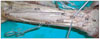
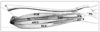
An AFCU was found unilaterally during routine dissection of a 65-year-old male cadaver. The anomalous muscle was located on the anteromedial aspect of the left forearm, in a plane anteromedial to FDS and between FCU and FDS, and was extended on the radial side of the FCU. The anomalous muscle belly (AFCU) originated from the medial epicondyle approxiamately 1 cm posterolateral to origin of normal FCU, and from proximal part of the flexor digitorum superficialis muscle. At proximal two thirds of anterior surface of the forearm, there were the musculotendinous connections between the fibres of AFCU and humero ulnar head of FDS, and also at 2 cm distal to origin of the FCU between the FCU and the AFCU. It formed an independent tendon 5 cm above the proximal edge of the flexor retinaculum.
On the ventral aspect of the hand, distal to the flexor retinaculum, the tendon of AFCU was located anterolateral to the unlar nerve, anteromedial to median nerve and medial to the tendons of FDS and just lateral to normal FCU. The AFCU inserts to the triquetral and hamate bones and flexor retinaculum. The fibres of abnormal muscle crossed the transverse axis of radiocarpal junction. Its tendon extended to carpal region and passed through the carpal tunnel. The abnormal muscle belly was measured 23.5 cm in length, 1.7 cm in width.
Passive traction on the tendon of accessory muscle resulted in flexion of the radiocarpal junction. The FCU which had one head arose from the medial epicondyle, and crossed from the anterior to Guyon canal, and inserted to the pisiform bone hook of hamate and palmar aponeurosis. The anomalous muscle on the left was innervated by a branch from the ulnar nerve, 2.6 cm distal the medial epicondyle (Figs. 1, 2). There was no other abnormalities than the AFCU.
Discussion
The neurovascular bundle of the forearm takes part in ulnar forearm pathway which is formed by FCU, FDS and Flexor digitorum profundus (FDP). It includes ulnar artery, vein and venae comitantes. To find the neurovascular bundle both in surgery and diagnostic techniques, the FCU is an important anatomical guideline and it is easly palpated in its distal course if the wrist is flexed and adducted [27].
Different anomalies of flexor muscles of forearm have also been described. The FDP may be joined by accessory slips from the radius, from FDS, FPL (Flexor pollicis longus), medial epicondyle or coronoid process of the ulna [16, 24, 26, 28]. A separate FDS tendon for the index finger has been identified [24]. Still and Kleinert [29] found an aberrant pollicis longus muscle (PL) passing through Guyon's canal. Jeffery [8] described an anomalous muscle in the lower forearm in the absence of PL refered as accessory abductor digiti minimi.
Williams et al. [26] reported that the tendon of abnormal muscle inserted on the proximal phalanx where as those of the FDS inserts on the middle phalanx. Sälgeback [30] identified an accessory muscle arising from the forearm fascia and flexing the metacarpophalangeal joint of the fifth digit. Pribyl and Moneim [31] found an accessory muscle originating from the FCU which joined flexor digiti minimi muscle in Guyon's canal. However, none of these muscles displayed attachments corresponding to the muscle described in our case.
Different origins and insertions for the AFCU have been described. The AFCU brevis which originates from the distal ulna may inserts into any of the following: pisiform, hook of the hamate, the abductor digiti minimi or the end of the fifth metacarpal [32]. The another abnormal muscle which named as ulnaris extensor brevis and originates from the medial epicondyle, inserts on the fourth and fifth metacarpals [16]. Nayak et al. [7] have described anomalous muscles which located on the ventral aspect of the proximal forearm in a plane deep to the FDS. In their cases, anomalous muscles originated from the deep surface of the FDS and it on the radial side inserted into the tendon of the flexor PL. In one of Kopuz's recent studies concerning an additional muscle in the forearm,the importance of these accessory muscle bellies or anomalous muscle slips has become apparent. It has been found that a separate muscle belly extending from the distal part of forearm into the carpal canal joins the tendon of flexor digitorum profundus muscle for the index finger [2].
In one case, it has been found that the anterior fibres of the FCU formed an accessory muscle which joined the main belly below the middle of the forearm [16]. Ang et al. [17] had found an accessory flexor muscle belly which running radial to the FCU proper, between FCU and FDS, and arising from the common flexor origin with its insertion at the triquetral bone .
Since the identified muscle in the present case was physically connected to forearm muscles it is similar to the above cases of other authors. However, the insertions of independent tendon of abnormal muscle does not correspond to any of the possible insertions of the anomalies described by other authors above except Anson's [32] and Ang et al.'s [17] ones. This difference needs to be embryologically explained.
The anatomical variation in the present case is a separate muscle belly extending to the hamate and triquetral bones and flexor retinaculum from medial epicondyle. The insertion of independent tendon of abnormal muscle coursing at the posterior to transverse carpal ligament similar to other flexor muscles or FCU having one head, and crossing from the anterior to Guyon canal here is interesting. This unusual muscle mimicking FCU may perhaps be beter named "musculus flexor carpi ulnaris accessorius or flexor carpi ulnaris palmaris" to denote their important relationship to the carpal bones, ulna or palmar aponeurosis.
Four fundamental phases have been described in the ontogenesis of muscle patterns [33]. The accessory muscle described here could have arisen during phase 3 [33] when muscle primordia from different layers fuse to form a single muscle [34]. However, stated that some muscle primordia disappear through cell death despite the fact that cells within them have differentiated to the point of containing myoflaments. Persistence of some cells between the FDS and FCU or persistence of some cells on FDS may account for the anomalous muscle slip in the case described here.
This abnormal or accessory muscle is not anatomically the only important. Abnormal muscle in this region can compress the unlar nerve within Guyon's canal [3]. It has been stated that the compression is rare at the level of the wrist [35, 36] and ulnar artery thrombosis may be associated with an anomalous muscle in Guyon's canal [31]. Also the diagnosis of an accessory muscle belly should be kept in mind when the mass is soft and in line with one of the fingers, and there is increased firmness an active contraction on the muscle against resistance [37]. Additionally knowledge of such variations supplements the anatomical information on the muscles of the antebrachial and carpal regions may become significant in preoperative diagnosis and in the hand during surgery. Therefore differences between frequencies of abnormal muscles of forearm and wrist in the further pediatric and adult series should be evaluated to understand the developmental patterns abnormal muscles.
 XML Download