

 PDF
PDF ePub
ePub Citation
 Print
Print


Introduction
Detailed surface three-dimensional (3D) models of the male urogenital system and related structures in the abdomen, pelvis, and perineum are useful for interactive medical simulations such as for virtual operations by urologists.
All surface models can be stereoscopically drawn by artists using anatomical knowledge, but satisfactory models can be easily acquired using a scientific approach. They seem to be obtained by digitalization of dissected male urogenital organs using a 3D scanner. However, dissecting each organ (e.g., ureter) with its original shape and location is impossible. Consequently, serial images are the only source to generate surface models, in which the structures are delineated and surface reconstructed. Computed tomography (CT) scans or magnetic resonance images (MRIs) are insufficient to show details of male urogenital organs and surrounding structures (e.g., corpus cavernosum and ischiocavernosus), even if contrast medium is injected. We believe that serially sectioned images of a cadaver, which contain actual anatomical color and high resolution, are preferable to CT scans or MRIs. Among sectioned images, the Visible Human Project male data show an absence of the right testis and a resolution limit (pixel size, 0.33 mm) [1], but the Visible Korean male data show the urogenital organs free from pathology with enhanced resolution (pixel size, 0.2 mm) [2]. Incidentally, unlike the Chinese Visible Human [3] and the Virtual Chinese Human [4], the Visible Korean cadaver has similar color to a living human, as no fixatives or dyes were injected, which is helpful to precisely identify various kinds of structures [5].
Computer language programming can be used for outlining and surface reconstruction; however, it is difficult to program software that satisfies every researcher. In this study, we set up a process to build surface models on widely available commercial software, so that the process can be reproduced by other investigators without the need for programming.
We have already experienced outlining of the male urogenital organs from the Visible Korean on SURFdriver ver. 3.56. However, the outlining procedure on SURFdriver was almost completely manual and was time consuming [6]. Therefore, we used Photoshop CS3 ver. 10 (Adobe Systems, Inc., San Jose, CA, USA), which is regarded as the best software for outlining sectioned color images [7]. However, stacked outlines of thin structures may not overlap on Photoshop, which would prevent automation of the next surface reconstruction procedure. Thus, we used the interpolation function on Discreet Combustion ver. 4 (Autodesk, Inc., San Rafael, CA, USA) to create overlap [8].
We also practiced surface reconstruction of urogenital organs using SURFdriver. However, the resultant surface models could only be opened on SURFdriver, and modifying the incorrect regions and reducing the polygons was impossible [6]. To solve this problem, we attempted surface reconstruction on Rhinoceros ver. 3.0 (McNeel North America, Seattle, WA, USA) and refined the surface models on Maya ver. 2009 (Autodesk, Inc.). Nevertheless, the results were unsatisfactory because some surface reconstruction procedures had to be performed manually, resulting in a long tedious job [9]. To save time and effort, surface reconstruction by way of volume reconstruction has been developed on AutoCAD ver. 10 (Autodesk, Inc.), but some manual procedures still remained for volume modeling [10]. Subsequently, it was recognized that 3D-DOCTOR ver. 4 (Able Software, Corp., Lexington, MA, USA) was useful for more automated volume reconstruction [11, 12].
Unlike volume models, surface models with a small file size can be manipulated in real time, but they are unable to show actual structure color [9]. Thus, surface models would be more realistic after being painted with the real surface texture of the structure. It is obvious that the more realistic the surface model, the more practical the medical simulation.
The objective of this research was to present realistic surface models of the male urogenital organs and neighboring structures made from the Visible Korean. Another purpose was to introduce the advanced surface reconstruction technique on popular software, which can be applied to create other useful surface models from serial images.
Materials and Methods
Serially sectioned images of a whole male cadaver (33 years old) had been acquired with 0.2 mm-sized intervals, 0.2 mm-sized pixels, and 24 bit color [5]. Among the whole body sectioned images, 464 images of the abdomen, pelvis, and perineum from the kidneys to the testis were selected at 1 mm intervals.
Outlining and outline interpolation
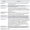
Male urogenital organs and other identifiable structures in the abdomen, pelvis, and perineum were chosen to be outlined and surface reconstructed (Table 1).
Pipe-like structures were outlined in three ways. The urinary bladder was outlined along both mural and luminal contours (Table 1) because the wall and the lumen were large and definite. But the ureter and urethra were outlined only along mural contours, as luminal contours were too narrow to be identified. Arteries and veins were outlined along their luminal contours, which were more distinct than mural contours.
The sectioned images (tag image file format, TIFF) were loaded into Photoshop. The "sharpen" filter was occasionally applied to make the structure border clearer, and the "median" filter was applied to generate the inside color of the structure with less noise. The structures were outlined automatically using the "magic wand selection" tool, semiautomatically using the "quick selection" or "magnetic lasso" tool, manually by "lasso" tool in sequence. Incorrectly drawn outlines were modified by moving the anchor and control points. The serial outlines of each structure were isolated and saved as outlined images of the structure (Photoshop documentary, PSD) [7].
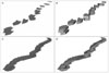
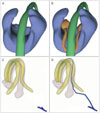
Neighboring outlines (1 mm intervals) were generally overlapped, but occasionally they were not overlapped in cases of thin structures such as the ductus deferens. The non-overlapping outlines produced several separate volume models and eventually disconnected surface models, which had to be connected manually. Therefore, after loading the neighboring outlined images (PSD files) onto Discreet Combustion, interpolation was conducted using the "animate" tool to generate intervening outlines, which overlapped with one another (Fig. 1).
Interpolation on Discreet Combustion was also employed to decrease the amount of outlining required. It was practically useful for structures that gradually changed along many serial sectioned images. For example, skin was outlined at 10 mm intervals; then interpolation was performed, creating 1 mm interval outlines. In cases in which the created outlines did not fit the structures in the sectioned images, the outlines were manually revised, which was much easier than outlining at 1 mm intervals.
Volume and surface reconstruction
After converting the file format of the outlined images from PSD to TIFF, black-filled outlines of a structure were sequentially accumulated on 3D-DOCTOR. For the proper horizontal-vertical proportion of the surface model, intervals of stacked outlines were determined based on the original intervals of the outlined images [9]. For example, if the intervals of the outlined images were narrowed by interpolation, intervals of the stacked outlines were adjusted closely.
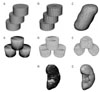
On 3D-DOCTOR, black-filled outlines were simultaneously extruded on the next outlines to create several small-volume 3D models, which comprised volume reconstruction. Subsequently, all volume models of the structures were combined (Figs. 1 and 2) [11, 12].
A surface 3D model was automatically extracted by covering the combined volume models on 3D-DOCTOR, which constituted the surface reconstruction. The surface model consisted of original stacked outlines and numerous polygons (planar triangles) between the outlines (Fig. 2). The surface model was exported into drawing exchange format (DXF) file.
Refining, assembling, and drawing of the surface models
Imperfections in each surface model (DXF file) were refined on Maya as follows. The original outlines and the stairs on the surface model were deleted using the "smooth" command. During this procedure, the number of polygons was unintentionally increased. The polygons were appropriately reduced in number using the "reduce" command without distorting the shape of the structures. As a result, the polygons were almost regular triangles (Fig. 2).
Flawed surface regions resulting from erroneous delineation were manually revised on Maya until the surface models corresponded to the actual anatomy.
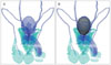
Collective outlined images (PSD files) of all structures were saved as Adobe Illustrator (AI) files. The outlines from the AI files were stacked with their proper locations retained in Maya [9]. The surface model of each structure, already built on 3D-DOCTOR and Maya, was manually placed to fit stacked outlines of the structure (Fig. 3). Then, the stacked outlines were deleted [12].
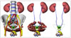
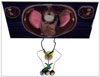
In a Maya file, the format of which is Maya binary (MB), layers were created and designated according to the structure names, which followed Terminologia Anatomica (Table 1) [13]. The surface model of each structure was then placed in its own layer to construct assembled surface models. The surface models were painted using "surface shader" to distinguish them (Fig. 4).
Small but significant structures (e.g., bulbourethral gland) were manually drawn on Maya to acquire additional surface models. The stereoscopic drawing was made using the "sphere" command and the "edit mesh" tool in association with surrounding structures. Similarly, thin but important structures of which only the proximal part was identifiable (e.g., dorsal artery of penis), were manually extended to prepare entire surface models (Table 1, Fig. 5) [12].
Coating of the surface model
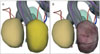
We composed software (Texture-extractor) in which the volume model of the testis was built from both sectioned images and outlined images of the testis, and surface texture of the testis was extracted from the volume model. Then, the surface model of the testis was coated with the surface texture on Maya (Fig. 6).
Results
In total, 95 surface models of urogenital organs and other structures were prepared, including complete urinary and genital tract models (Table 1). The surface models, shown in their original location, reflected the proper shape of the structures (Fig. 4). Many surface models could be objectively built with automation, as a new interpolation technique on Discreet Combustion (Fig. 1) and 3D reconstruction on 3D-DOCTOR (Fig. 2) were developed and applied.
The surface models comprising only surface polygons had small file sizes in contrast to the volume models. The optimized surface models, with the polygons reduced (Fig. 2), could be handled in real time, suggesting their use for interactive medical training simulation.
The surface models in an MB file could be conveniently viewed in many ways on Maya. Any surface model combination could be selected for display using the "layer" window. The surface models could be shaded for stereoscopic effect using the "hypershade" tool. Additionally, the color of individual surface models could be changed using the "color" tool. We could make the color semitransparent so that we could see behind the surface models. Surface model composites could be rotated at arbitrary angles (Fig. 4). Additionally, we found that we could produce a surface model with the actual texture of the human organ (Fig. 5). The texture effect was enhanced in the present study because the cadaver was serially sectioned without fixative or dye injections [5].
Discussion
Outlining, surface reconstruction, refining, and assembling are necessary to obtain surface models from serial images. In some studies, all procedures were performed in a single software application such as SURFdriver [6] or 3D-DOCTOR [14, 15]. The problem with this is that these applications were originally developed for 3D reconstruction, so other procedures (outlining, refining, etc.) are not automated. In this study, we combined the use of popular software (Photoshop, Discreet Combustion, 3D-DOCTOR, and Maya) to increase the automation of all steps. The images were transferred between the different software applications: TIFF files between Photoshop and 3D-DOCTOR, DXF files between 3D-DOCTOR and Maya.
We improved the technique by trial and error to determine the appropriate commands in the software. From our experience, the following systemic principles are strongly recommended to obtain surface models and to save time and effort.
Surface reconstruction should be accomplished after volume reconstruction. Surface reconstruction to fill gaps between piled outlines with polygons cannot be done automatically in the case of dividing structures such as an arterial branch [9]. This problem is solved by using volume models: Volume reconstruction of stacked outlines produces several small volume models, from which a surface model can be extracted regardless of the dividing regions (Fig. 2) [10].
Both volume and surface reconstruction can be achieved on 3D-DOCTOR. We have used AutoCAD for volume and surface modeling [10]. On AutoCAD, several commands including "filling outlines," "extruding," and "extracting surface" are essential in successive steps; but on 3D-DOCTOR, fewer commands are sufficient for the same reconstruction. This is because AutoCAD is a software with numerous functions for multiple purposes, whereas 3D-DOCTOR is software specifically for 3D serial image modeling [11, 12].
Volume and surface reconstruction is executed after interpolation on Discreet Combustion (Fig. 1). Initially, interpolation seemed to require programming; but we found a tool on Discreet Combustion that enabled interpolation without depending on programming [8]. Instead, we expect that programmers will develop an educational program from our surface 3D models.
Surface models of different structures are assembled on proper locations on Maya. A drawback of 3D-DOCTOR is that the reconstructed surface models have no information about location. Thus, information was derived from the stacked outlines of all structures (Fig. 3). As a result, end users are provided with the accurately assembled surface models (Fig. 4) [12].
Additional surface models can be manually drawn on Maya. The important structures, which are hardly identified in the sectioned images, need to be drawn manually. For example, the bulbourethral glands were drawn referring to anatomy knowledge: The two pea-size bulbourethral glands lie posterolateral to the intermediate part of the urethra, and are largely embedded within the external urethral sphincter (Table 1, Fig. 5). This scientific drawing based on objective surface models is considerably different from an artist's drawing [12].
The surface model is coated by real surface texture using Texture-extractor and Maya. We attempted to acquire the surface texture for the volume model on Texture-extractor that was the exceptional programmed software in this research. The problem was that outlining was not perfect, so consistent real surface texture could not be obtained (Fig. 6). To solve this problem, Texture-extractor must be upgraded as follows. A volume model is superimposed by a surface model of a structure, and the volume model is transparent except for the voxels encountered with the surface model. The visualized voxels are the surface texture of the structure. An incorrect surface cause the surface texture to show an inconsistent color. Some regions of the surface model were interactively expanded or shrunk until the surface texture became consistent. This procedure might result in a revised version of the outlined images.
We utilized popular commercial software packages such as Photoshop, Discreet Combustion, 3D-DOCTOR, and Maya, because their availability will allow other investigators to perform outlining and surface reconstruction on a personal computer without requiring the assistance of computer engineers. Using these packages, investigators can take advantage of user friendly commands and obviate debugging procedures to produce surface models from sectioned images, CTs, and MRIs.
The surface models prepared during this study are available free of charge. After obtaining permission to use data from our group, data will be provided to users worldwide either on or off-line. The surface models are distributed together with the sectioned and outlined images, all of which correspond to one another. Additionally, the equivalent CTs and MRIs of the same cadaver can also be presented to enhance the value of the surface models [5]. Beneficial software can be produced from the various kinds of images that complement each other. For example, surface models are superimposed on the sectioned images for supplementary comprehension of the structures (Fig. 7).
Almost 100 surface models from this investigation are expected to be applied for a variety of purposes [16, 17]. For example, surface models of the perineum could be a source for a virtual endoscopic prostatectomy for urologists; surface models of the urinary system could be a source of a learning program about urinary continence and incontinence for medical students; surface models of the male genital system could be the source of either an animation for sperm passage or an interactive simulation game for contraception and vasectomy.
 XML Download