

 PDF
PDF ePub
ePub Citation
 Print
Print


Introduction
Excessive calcium (Ca2+) is thought to be a critical step in various neurodegenerative processes including ischemia. During cerebral ischemia, a series of biochemical events occur that causes depolarization, activation of voltage-gated Ca2+-channels and an excessive release of excitatory amino acids, especially glutamate. Activation of glutamate receptors leads to an increase in intracellular Ca2+ and the activation of Ca2+-dependent catabolic enzymes such as phospholipases, protein kinases, proteases, and caspase cascades (White et al., 2000), which subsequently induce neuronal cell death. Past exploration of this complex pathophysiology has resulted in the development of a large number of candidates for neuroprotective intervention. It is noteworthy that a number of Ca2+-channel blockers have been developed and tested for use in ischemic stroke treatment. However, they have failed in clinical trials because of either a lack of therapeutic efficacy or drastic side effects (Azcona & Lataste, 1990; Limburg & Hijdra, 1990; Rosenbaum et al., 1991; Ahmed et al., 1996).
Calcium binding proteins (CaBPs) are thought to play a major role in buffering intracellular Ca2+ and to be involved in a variety of Ca2+-mediated signal transduction (Rogers 1987; Polans et al., 1996; Schäfer & Heizmann, 1996). Three CaBPs, namely, calbindin D28k (CB), parvalbumin (PV) and calretinin (CR), members of the EF-hand calcium-binding protein family, have been implicated to play a neuroprotective role in various pathologic conditions by functioning as buffers for excessive calcium. In particular, CB is found in neurons throughout the central nervous system (Celio et al., 1990; Baimbridge et al., 1992; Lewit-Bentley & Réty, 2000) and reactive astrocytes in some pathologic conditions (Toyoshima et al., 1996; Lee et al., 2004), and has the ability to buffer and modulate intracellular Ca2+ concentrations (Batini et al., 1993 and 1997). In addition, it has been reported that exogenous CB can reduce oxidative stress, preserve mitochondrial function and protect neurons against glutamate- or ischemia-induced excitotoxicity (Guo et al., 1998; Meier et al., 1998; D'Orlando et al., 2001 and 2002). However, relatively limited information concerning the neuroprotective function of CR and PV is available.
Although there are some differences in expression patterns among the species, CaBPs are expressed in specific neurons in the mammalian retina, as in other regions of the brain. Thus, anti-CaBPs have been used as markers for specific neuronal populations and/or subpopulations. In the rat retina, anti-CB can be used to label horizontal cell population and subpopulations of amacrine and ganglion cells (Pasteels et al., 1990; Chun et al., 1999). CR and PV are used to label different subsets of amacrine and ganglion cells (Pasteels et al., 1990; Sanna et al., 1990; Mojumder et al., 2008; Park et al., 2008). Using differential labeling with anti-CaBPs, specific retinal neurons have been characterized in both normal and various pathologic conditions.
Retinal ischemia induced by increasing intraocular pressure has been widely used to examine the effects of ischemic damage, which leads to cellular changes in a variety of bioactive molecules in the retina including neurotransmitters and CaBPs, as well as changes in retinal neuronal populations or subpopulations. Using the real-time quantitative polymerase chain reaction (PCR) (Dijk et al., 2004) and immunohistochemistry (Dijk & Kamphuis, 2004), Kamphuis and co-workers reported that the transcript levels of PV and CR were decreased and the number of each expressing amacrine subpopulation was substantially reduced in the rat retina after ischemia/reperfusion insults. Using immunohistochemistry, Kwon et al. (2005) reported that CB-expressing ganglion cells were more resistant to ischemic damage than CR- and PV-expressing ganglion cells in the rabbit retina. These results suggest that CaBPs appear to be related to a differential vulnerability among retinal neurons to ischemic damage probably due to their different Ca2+ buffering abilities. At this point, in order to confirm previously reported results and to extend our knowledge related to the neuroprotective function of CaBPs and the vulnerability of retinal neurons to ischemic insult in the retina, we investigated changes in the expression of three calcium binding proteins, CB, PV and CR in ischemic rat retina induced by increasing intraocular pressure, at the transcript and protein levels using quantitative real-time reverse transcription (RT)-PCR, western blot and immunohistochemistry.
Materials and Methods
Animals
Thirty-five adult female Sprague-Dawley rats weighing 200~250 g were used in this study. The animals were treated according to the regulations of the Catholic Ethics Committee of the Catholic University of Korea, Seoul, which conform to the National Institute of Health (NIH) guidelines for the Care and Use of Laboratory Animals (NIH Publications No. 80-23) revised 1996.
Induction of transient retinal ischemia and tissue preparation
Animals were anesthetized with 4% chloral hydrate (1 mL/100 g body weight). The pupils were dilated with 1% tropicamide drops. The intraocular pressure (IOP) was increased to 90~120 mm Hg by cannulation of the anterior chamber with a 26-gauge needle connected to a hydrostatic pressure device, and the elevated IOP was maintained for 60 min. The applied pressure of 90~120 mm Hg is in the range or slightly above the systemic systolic pressure in the normal rat (Büchi et al., 1991). Following removal of the cannula, recirculation was immediately initiated and the IOP decreased to normal values within 5 min.
Animals were sacrificed by an overdose of chloral hydrate at different time points after reperfusion: 1, 3 days, 1, 2, 4, 8 weeks (wks). The anterior segments of the eyeballs were removed. For real-time PCR and western blot analysis, retinal tissues were quickly dissected on an ice-cold plate, frozen on dry ice, and stored at -70℃. For immunohistochemistry, the eyecups were fixed by immersion in 4% paraformaldehyde in 0.1 M phosphate buffer (PB, pH 7.4), for 2~3 h. Following fixation, the retinae were carefully dissected and transferred to 30% sucrose in PB, for 24 h at 4℃. They were then frozen in liquid nitrogen, thawed, and rinsed in 0.01 M phosphate-buffered saline (PBS, pH 7.4).
Isolation of total RNA
Frozen retinas were homogenized and then suspended in 1 mL of TRIZOL reagent (Invitrogen, Calrsbad, CA). The homogenized samples were incubated for 5 min at room temperature and 0.2 mL of chloroform was added to each sample. The sample tubes were shaken vigorously by hand for 15 seconds and then incubated at room temperature for 5 minutes. The mixture was separated into a lower red, phenol-chloroform phase, an interphase, and a colorless upper aqueous phase by centrifugation at 13,000 rpm, 4℃ for 15 minutes. The aqueous phase was transferred to a new tube and 0.5 mL of isopropyl alcohol added. After incubation for 10 minutes at room temperature, the samples were centrifuged at 13,000 rpm, 4℃ for 15 minutes. The RNA precipitate formed a gel-like pellet on the side and bottom of the tube. The RNA pellet was washed once with 75% ethanol, then air-dried and re-dissolved in 30 µL 0.1% DEPC water. Total RNA was quantitatively measured with UV/Vis spetrophotometry (ND 1,000, Nanodrop, Wilmington, USA).
Reverse transcription
A two microgram sample of total RNA from retinal tissues was reverse transcribed using a High Capacity RNA-to-cDNA Kit (Applied Biosystems, Foster City, CA) according to the manufacturer's protocol. RNA template and 2X RT buffer and 20X Enzyme mix were incubated at 37℃ for 60 minutes, followed by heating at 95℃ for 5 minutes to stop the reaction, after which, the cDNA products were stored at 4℃.
Quantitative real-time RT-PCR
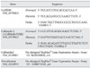
Real-time RT-PCR was performed using a Rotor-Gene 6000 System instrument and software (Corbett Research, NSW, Australia). The relative expression level of one housekeeping gene and three target genes was measured by the TaqMan® Gene expression-based method. For the real-time PCR analyses, we acquired gene-specific FAM dye-labeled primer-probe pairs: TaqMan MGB probes (Applied Biosystems). In this case, the exact primer and probe sequences were not available from the manufacturer. The sequences and information on the primers are presented in Table 1, and the specificity of the primers was confirmed by a BLAST search.
PCR reactions were prepared in a final volume of 20 µL, with 2X TaqMan® Gene expression Master mix (Applied Biosystems) and cDNA derived from 10 ng of input RNA on ice.
The cycling conditions were as follows: AmpliTaq activation at 95℃ for 10 minutes (one cycle), denaturation at 95℃ for 15 seconds and annealing/extension at 60℃ for 1 minute (50 cycles). All assays included a positive control and a no-template control. Each measurement was performed in duplicate and the threshold cycle (CT) was determined.
Statistical analysis
The CT value was determined per gene for each sample. The CT values were transformed to linear scale raw expression quantities using the Δ-CT method, and were calculated using the Excel software program. To compare gene expression between samples, the CT was normalized to an endogenous reference (GAPDH) and relative to a calibrator. Correlation of gene-expression analyses was performed by the Pearson linear correlation. P-values were considered to be significant if <0.05.
Western blot analysis
Western blot analyses were performed on retinal extracts that had been homogenized in 10 vol of 20 mM Tris-HCl, pH 7.4, 150 mM NaCl, 1 mM EDTA, 1% Triton X-100, 0.5% sodium deoxycholate, 0.1% sodium dodeyl sulfate (SDS), 0.02% sodium azide, 1 mM PMSF, and 5 µg/mL leupeptin. Protein concentration in each sample was assayed by the Lowry method (Lowry et al., 1951; Peterson 1979) in duplicate and the results averaged. Duplicate sets of protein standards containing 0, 1, 3, 5, 10, 20, 40, or 60 µg bovine serum albumin (BSA) were assayed by the same method. The results were averaged and graphed to produce a linear equation that was used to estimate the protein contents of the retinal extracts. The optical density of each sample was measured at 660 nm, using a spectrometer (Spectronic 20; Bausch and Lomb, Rochester, NY). Aliquots of tissue samples corresponding to 25 µg of total protein were heated at 100℃ for 10 min with an equivalent volume of 2× sample buffer (containing 4% SDS and 10% mercaptoethanol) and loaded onto 12% polyacrylamide gels. The proteins were electrotransferred to a nitrocellulose membrane in Tris-glycine-methanol buffer. The membrane was blocked for 1 h at room temperature in a blocking solution containing 5% nonfat dry milk, 0.1% Tween-20, and PBS (pH 7.4). The membrane was then incubated for 15 h at 4℃ with primary antibodies in the blocking solution. The following primary antibodies were used in the western blot experiments; mouse monoclonal anti-CB (Sigma, St. Louis, MO; dilution 1 : 10,000), rabbit polyclonal anti-CR (Millipore, Bedford, MA; dilution 1 : 100,000), mouse monoclonal anti-PV (Sigma; dilution 1 : 1,000), and mouse monoclonal anti-β-actin (Sigma; dilution 1 : 100,000) for a control. The membrane was rinsed three times with 0.05% Tween-20 in PBS for 10 min each, followed by incubation for 1 h at room temperature in a 1 : 200 dilution of biotinylated donkey anti-mouse IgG or anti-rabbit IgG (Vector Laboratories, Burlingame, CA). The blot was washed three times for 10 min each and then processed for analysis using an enhanced chemiluminescence (ECL) detection kit (Amersham, Arlington Heights, IL).
Immunohistochemistry
Retinal tissues were blocked in 10% donkey serum and incubated with primary and secondary antibody dilutions, prepared in 1% donkey serum in PB with 0.5% Triton X 100. Primary antibodies were incubated at 4℃ for 5~7 d, and secondary antibodies were incubated at 4℃ overnight. The antibodies were applied in immunohistochemistry, as follows; mouse monoclonal anti-CB (Sigma; dilution 1 : 1,000) and rabbit polyclonal anti-CB (Swant, Bellinzona, Switzerland; dilution 1 : 2,000), rabbit polyclonal anti-CR (Millipore; dilution 1 : 2,000), and mouse monoclonal anti-PV (Sigma; dilution 1 : 1,000). The tissues were washed in PB for 45 min (3×15 min), incubated in the presence of Cy3-conjugated donkey anti-mouse IgG or donkey anti-rabbit IgG (Jackson Immuno Research, West Grove, PA; dilution 1 : 100) for 2 h. After thoroughly washing with PB, the retinal tissues were mounted with Vectashiled H-1000 (Vector Laboratories).
Digital images (1,024×1,024 pixels) were acquired using a Zeiss LSM 510 Meta confocal microscope (Carl Zeiss Co., Ltd., Germany). Images were converted into TIFF format, and processed for contrast level adjustment using Photoshop v. 7.0 (Adobe Systems, San Jose, CA).
Cell number counting
The number of the labeled cells was counted in five retinal wholemount peices of each group using an image analysis system Image-Pro Plus (I-CUBE, Glen Burnie, MD) and represented as the density (given in cells/mm2). Using a 20× objective and 10× eyepieces, images of 10 sample fields per retina were captured and then processed. The density is described as mean±SD.
Results
Changes of CB, CR and PV transcript levels
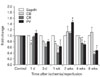
To examine changes in the expression of the three calcium binding proteins, CB, CR, and PV, in ischemic rat retina induced by increasing intraocular pressure, quantitative real-time RT-PCR was performed at each time point. Following ischemia/reperfusion, changes in CB, CR and PV transcripts levels showed differential patterns (Fig. 1).
CB transcript levels showed a reduction (on average 75%) from 1 day to 1 wk after ischemia/reperfusion; thereafter, the levels fluctuated. At 2 wks, a maximum was reached at 114% of the control level, the levels reverted to 93% at 4 wks, and at 8 wks, the level had increased to 112%. However, these changes during the latter half of the experimental period was statistically insignificant (Fig. 1, P>0.05).
Following ischemia/reperfusion, a gradual decrease in CR transcript levels was observed from 1 day onward; at 1 wk, the value reached 42%. At 2 wks, the level rebounded and increased to 110%; thereafter the levels continued to decrease, finally reaching 37% of the control transcript level remaining (Fig. 1, P<0.05).
The pattern of changes in transcript levels for PV was quite similar to that of CR (Fig. 1). Two differences were observed. One was an initial increase in PV transcript. The level reached 108% of the control level, but was statistically insignificant (P>0.05). The other change was a significant peak at 2 wks (P<0.05). In the long term (4 and 8 wks after ischemia/reperfusion), the transcript levels were significantly lower by 71% and 50% of the control transcript level remaining (Fig. 1, P<0. 05).
Changes of CB, CR and PV protein levels by Western blot
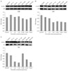
The expression of CB, CR, and PV proteins was easily detectable in control retinas (Fig. 2). CB, CR and PV were detected as a single band at a 28-kDa, 29-kDa, and 12-kDa band, respectively. Following ischemia/reperfusion, the expression patterns of CB, CR and PV proteins were quite different among the three proteins.
Compared to the control level, the levels of the CB protein did not changed significantly in response to ischemia/reperfusion throughout the experimental period (Fig. 2A).
The level of expression of CR began to decrease by 1 wk after ischemia/reperfusion and continued to be low thereafter. At 8 wks after ischemia/reperfusion, the protein level was significantly lower at 54% of the control level remaining (Fig. 2B).
In response to ischemia/reperfusion, the levels of PV protein rapidly decreased by 1 wk and reached a minimum of 29% of the control level. Thereafter, the expression recovered, reaching 97% at 2 wks. Afterwards, the PV protein levels again rapidly decreased, reaching 32% of the control level at 8 wks.
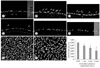
Changes in CB, CR and PV-expressing retinal neurons by immunohistochemistry
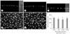
Although CB is distributed in horizontal, amacrine and ganglion cells in the rat retina, it is strongly expressed in horizontal cells and thus has been used as a horizontal cell marker (Pasteels et al., 1990; Chun et al., 1999). In vertical sections of control retina (Fig. 3A), strong CB immunoreactivities were found in horizontal cell somata in the outer part of the inner nuclear layer (INL) and their dendrites in the outer plexiform layer (OPL) and very faint immunoreactivities were observed in the inner part of the INL, inner plexiform layer (INL), and ganglion cell layer (GCL). These labeling pattern and number of CB-expressing cells appeared to be unchanged throughout the experimental period at a glance (Figs. 3B and C). These findings were clearly demonstrated in retinal whole mount preparations (Figs. 3D and E) and wereconfirmed by counting the CB-expressing horizontal cells (Fig. 3F).
CR immunoreactivities were found in numerous amacrine cells in the inner part of the INL and numerous cells in the GCL, and three bands in the IPL in the control retina (Fig. 4A). The number of CR-expressing cells located in both the INL and GCL decreased with passing time (Figs. 4B and C). In particular, a marked decrease was observed in cells in the GCL (Figs. 4D and E). Compared to the cells of the GCL in the control retina, 67% were present in the retina at 4 wks after ischemia/reperfusion injury, and only 27% remained at 8 wks (Fig. 4F).
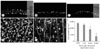
In the rat retina, PV is mainly expressed in AII amacrine cells in the INL and ganglion cells in the GCL in the minority, and thus anti-PV has been used as a marker for AII amacrine cells (Chun et al., 1993; Wässle et al., 1993). In the control retina, PV immunoreactivities were found in AII amacrine cells in the inner part of the INL and several ganglion cells in the GCL, and AII processes in the IPL (Fig. 5A). The numbers of PV-expressing AII amacrine cells and ganglion cells were gradually decreased (Figs. 5B~F). In retinal wholemounts at 8 wks after ischemia/reperfusion injury (Fig. 5H), 56% remained (Fig. 5I), compared to PV-expressing AII amacrine cells in the control retina (Fig. 5G). Interestingly, a new PV-expressing population, presumably cone bipolar cells was transiently observed in the outer part of the INL. These cells were found between 3 days and 2 wks after ischemia/reperfusion injury (Figs. 5B~D), with a peak at 3 days (Fig. 5B).
Discussion
Retinal ischemia/reperfusion can be a feature of pathologies such as amaurosis fugax and acute angle-closure glaucoma, and animal models have been used to study the mechanisms and effects of ischemic damage in the retina (Büchi et al., 1991; Kim et al., 1998; Li et al., 2009). Excessive Ca2+ is thought to be an important trigger, ultimately leading to ischemic neuronal degeneration (Kristián et al., 1998). CaBPs are thought to play a neuroprotective role in many pathologic conditions leading to neurodegeneration, by buffering excessive Ca2+. Thus, an impaired regulation of calcium by CaBPs is thought to be closely related to many neurodegenerative processes (Heizmann & Braun, 1992; Polans et al., 1996; Schäfer & Heizmann, 1996). Only a few studies have been reported dealing with changes in CaBPs after ischemia/reperfusion injury in the retina. However, most of these studies used CaBPs to identify specific retinal neuronal populations in a simple manner and the major focus was on the differential effects of ischemia/reperfusion injury on various retinal neuronal populations (Dijk et al., 2004; Dijk & Kamphuis, 2004; Kwon et al., 2005). In addition, the duration of the observations was relatively short (4 wks after ischemia/reperfusion injury). In the present study, therefore we examined the changes in both transcripts and proteins of three EF-hand CaBPs, CB, CR and PV, which are abundantly expressed in specific neurons of the mammalian retina, over a long period of time (Pasteels et al., 1990; Sanna et al., 1990; Wässle et al., 1993; Chun et al., 1999; Mojumder et al., 2008; Park et al., 2008), in rat retina induced ischemia/reperfusion injury produced by raising the intraocular pressure.
After ischemia/reperfusion retinal neurons in the GCL and INL undergo cell death via necrosis and apoptosis, and subsequently photoreceptors in the ONL (Büchi 1992a and 1992b; Choi 1996 and 2001; Kim et al., 2004). Despite the marked loss of retinal neurons, CB transcript and protein levels were not significantly changed in response to ischemia/reperfusion throughout the entire experimental period in this study. In addition, most of the CB-expressing horizontal cells had survived at the end of experimental period. These results are consistent with our previous immunohistochemical study (Chun et al., 1999) showing that horizontal cells labeled with anti-CB are resistant to degenerative processes to ischemic damage in the rat retina. These conclusions were also verified by Kwon et al. (2005) who reported that CB-immunnoreactive cells in the GCL of the rabbit retina were less vulnerable than CR- or PV-immunoreactive neurons. The expression of CB was increased in transient forebrain ischemia (Hwang et al., 2003) and spinal ischemia (Lee et al., 2005), which appeared to be related to resistance to neuronal degeneration. An over-expression of CB protected neuronal cells from signals that increase intracellular Ca2+ and induce apoptosis and thus could decrease neuronal death after transient focal ischemia (Yenari et al., 2001). In addition, Fan et al. (2007) recently demonstrated that CB protein transduction can significantly decrease neuronal death in cultures and an animal model of cerebral ischemia. Collectively, these findings indicate that CB might play a role in neuroprotection to ischemia/reperfusion injury in the retina and CB-expressing horizontal cells appear to have such resistance.
In contrast to CB, transcripts and proteins of CR and PV were decreased after ischemia/reperfusion in this study. At 8 wks after ischemia/reperfusion, the numbers of CR-expressing cells in the GCL and PV-expressing cells were only 27% and 56% of the control group, respectively. These results are consistent with previous reports by Kamphuis and co-workers (Dijk et al., 2004; Dijk & Kamphuis, 2004) who reported that transcripts and expressing cell numbers of CR and PV were decreased at 4 wk after ischemia/reperfusion. In terms of vulnerabilities of CR and PV in response to ischemia/reperfusion, however, our results were in conflict with their reports. In this study, among the three CaBPs, CR-expressing cells appeared to be the most vulnerable, while PV-expressing cells were identified as the most vulnerable ones in their study (Dijk & Kamphuis, 2004). This discrepancy might be caused by the differences in the ischemia/reperfusion injury model. That is, Dijk and Kamphuis used Wistar rats and induced retinal ischemia by increased intraocular pressure (IOP) using a 30-gauge needle connected to a saline reservoir, elevated to 1.7 m (Dijk & Kamphuis, 2004), while we used Sprague-Dawley rats and induced ischemia by increasing IOP using a 26-gauge needle connected to a hydrostatic pressure device, raised to 90~120 mm Hg. Although the difference in ischemia/reperfusion injury model appears to be minor, it should be recalled that a minor difference can cause quite different ischemic insults. For example, one of the cerebral ischemia models, the four vessel occlusion model using Wistar rats causes white matter degeneration in the brain, while that using Sprague-Dawley rats show hippocampal degeneration (Kim et al., 2008). Actually, the INL, IPL, and GCL were distinguishable at 4 and 8 wks after ischemia/reperfusion in this study (Figs. 3~5), while in the report by Dijk and Kamphuis (Dijk & Kamphuis, 2004), the IPL already appeared markedly thinned at 72 h and thus at 1 wk or 2 wks the IPL appeared a sheet. These results indicate that the ischemia/reperfusion injury in our model is milder than that of Dijk and Kamphuis (Dijk & Kamphuis, 2004) and at least in our retinal ischemia/reperfusion injury model, CR-expressing cells might be the most vulnerable.
Interestingly, a great decrease in CR-expressing ganglion cells occurred near the end of the study period, that is, from 67% of control at 4 wks to 27% at 8 wks (Fig. 4F). Considering that the retinal ganglion cell is the most vulnerable cell type in some pathologic conditions, such as glaucoma, retinal ischemia, and NMDA toxicity (Perlman et al., 1996; Adachi et al., 1998; Joo et al., 1999; Kageyama et al., 2000), our results suggest that CR may act as a Ca2+ buffer in CR-expressing ganglion cells, preventing them from degeneration and contribute to their survival from initial ischemia/reperfusion injury during the first half of an experimental period. In contrast to CR-expressing ganglion cells, PV-expressing AII amacrine cells were more decreased in the first half of the experimental period (27% reduction by 4 wks) than in the latter half (17% reduction) (Fig. 5F). CaBPs are thought to be involved in various Ca2+-mediated signal transduction processes in the central nervous system, in addition to buffering intracellular Ca2+ (Rogers 1987; Polans et al., 1996; Schäfer & Heizmann, 1996). Although the functions of other CaBP, including PV in the retina, remain unclear, our results suggest that PV may act as a mediator that is involved in some currently not understood Ca2+-mediated signal transduction within PV-expressing AII amacrine cells rather than acting as a Ca2+ buffer. The functions of CaBPs including PV need to be elucidated through the further studies.
Another interesting finding of this study is that a new PV-expressing neuronal population transiently appeared during the period 3 days to 2 wks. In the rat retina, PV is mostly highly expressed in AII amacrine cells, a crucial interneuron in the rod pathway, in the INL and thus, anti-PV has been used as a marker for AII amacrine cells (Chun et al., 1993; Wässle et al., 1993). These AII amacrine cells make extensive gap junctions with some types of ON cone bipolar cells (Massey & Mills, 1996 and 1999). Gap junctions generally transport molecules with molecular weights of below 1 kD in physiological conditions (Flagg-Newton et al., 1979). However, it has been demonstrated that the permeability of this gap junction can be altered in response to modulators such as dopamine and nitric oxide (Vaney et al., 1998; Xia & Mills, 2004). In addition, Park et al. (2008) recently reported the same phenomenon, in that a new group of PV-expressing cells was transiently found in the outer part of the INL in the rat retina experimentally induced diabetes and, using double-labeling technique, they demonstrated that they were the type 6 ON cone bipolar cells. Although we did not characterize the new population of PV-expressing cells in this study, therefore, it appears likely that this new population of PV-expressing cells in the INL are a certain type of ON cone bipolar cells and this transient expression of PV in some cone bipolar cells might be derived from an altered permeability, caused by ischemia/reperfusion injury, and not by de novo synthesis.
In conclusion, CB among the three CaBPs studied in this study, may have a neuroprotective function to ischemia/reperfusion injury through its powerful ability to buffer increased Ca2+ in the rat retina, and CR may provide some resistance to ischemia/reperfusion to CR-expressing ganglion cells but not as much as CB. In addition, PV may mainly act as a mediator involved in some type of Ca2+-mediated signal transduction within PV-expressing AII amacrine cells, rather than as a Ca2+ buffer.
 XML Download