Abstract
Neonatal diaphragmatic hemangioma (NDH) with diffuse neonatal hemangiomatosis has rare incidence. Hydrops fetalis can be found on prenatal ultrasonography and pleural effusion, pericardial effusion, and ascites can be found on postnatal ultrasonography. NDH can be treated with medical, interventional and surgical methods. We report a rare case of NDH which was successfully treated by coil embolization. The patient presented with cardiac tamponade, respiratory distress and abdominal distension are observed after birth due to massive fluid production by diaphragmatic hemangioma. Two times of interventional coil-embolization has made the infant's symptoms and signs alleviated through decreasing massive fluid collected in pericardial, pleural and abdominal space.
REFERENCES
1). Mandal AK., Lee H., Salem F. Review of primary tumors of the diaphragm. J Natl Med Assoc. 1988. 80:214–7.
3). Cacciaguerra S., Vasta G., Benedetto AG., Bagnara V., Guarnera S., Bartoloni G, et al. Neonatal diaphragmatic hemangioma. J Pediatr Surg. 2001. 36:E21.

4). Wu L., Wang JM., Qiao ZW., Yan YL., Wang LS. Successful embolization and long-term follow-up of a rare neonatal diaphragmatic hemangioma. SAGE Open Med Case Rep. 2015. 3:2050313X15615471.

5). Kaniklides C., Dimopoulos PA. Diaphragmatic haemangioma. A case report. Acta Radiol. 1999. 40:329–32.
6). Cambonie G., Saguintaah M., Masson F., Prodhomme O., Boulot P., Couture A, et al. Rapidly involuting congenital diaphragmatic hemangioma. Eur J Radiol Extra. 2009. 72:e125–8.

7). Olsen L., Gustafsson G., Kreuger A., Pech P. Un unusual case of hemangioma of the left diaphragm in a child. Successful use of Lyodura S for repair of the diaphragmatic defect. Pediatr Surg Int. 1995. 10:259–60.

8). George A., Mani V., Noufal A. Update on the classification of hemangioma. J Oral Maxillofac Pathol. 2014. 18(Suppl 1):S117–20.

9). Matulich J., Wood G., Sugo E. Case of non-involuting congenital haemangioma. Australas J Dermatol. 2005. 46:165–8.

10). Luu M., Frieden IJ. Haemangioma: clinical course, complications and management. Br J Dermatol. 2013. 169:20–30.

11). Restrepo R., Palani R., Cervantes LF., Duarte AM., Amjad I., Altman NR. Hemangiomas revisited: the useful, the unusual and the new. Part 1: overview and clinical and imaging characteristics. Pediatr Radiol. 2011. 41:895–904.
12). Krol A., MacArthur CJ. Congenital hemangiomas: rapidly involuting and noninvoluting congenital hemangiomas. Arch Facial Plast Surg. 2005. 7:307–11.
13). Liang MG., Frieden IJ. Infantile and congenital hemangiomas. Semin Pediatr Surg. 2014. 23:162–7.

14). Tsang FH., Lun KS., Cheng LC. Hemangioma of the diaphragm presenting with cardiac tamponade. J Card Surg. 2011. 26:620–3.

15). Curros F., Brunelle F. Prenatal thoracoabdominal tumor mimicking pulmonary sequestration: a diagnosis dilemma. Eur Radiol. 2001. 11:167–70.

16). Bellini C., Hennekam RC. Non-immune hydrops fetalis: a short review of etiology and pathophysiology. Am J Med Genet A. 2012. 158A:597–605.

17). Ismail KM., Martin WL., Ghosh S., Whittle MJ., Kilby MD. Etiology and outcome of hydrops fetalis. J Matern Fetal Med. 2001. 10:175–81.

18). Bellini C., Hennekam RC., Fulcheri E., Rutigliani M., Morcaldi G., Boccardo F, et al. Etiology of nonimmune hydrops fetalis: a systematic review. Am J Med Genet A. 2009. 149A:844–51.

Fig. 1
Radiograph showed a mass in the right lower hemithorax. Bilateral pleural effusion and ascites were observed.
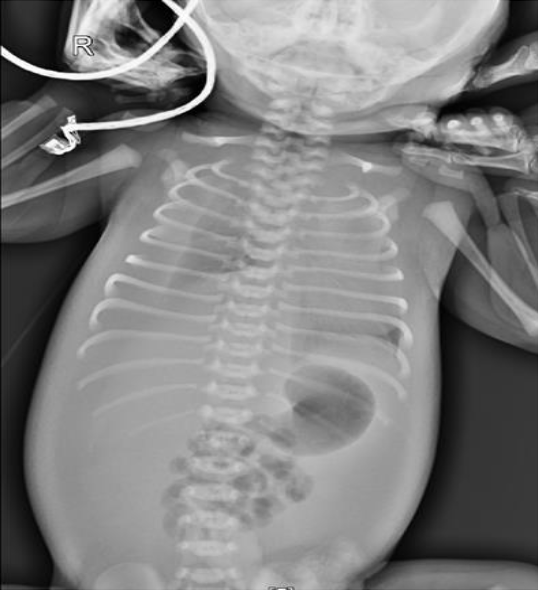
Fig. 2
Chest computed tomography axial image (A), coronal image (B) showed 5×5×2 cm sized, lobulated, multiseptated enhancing mass between heart and diaphragm.
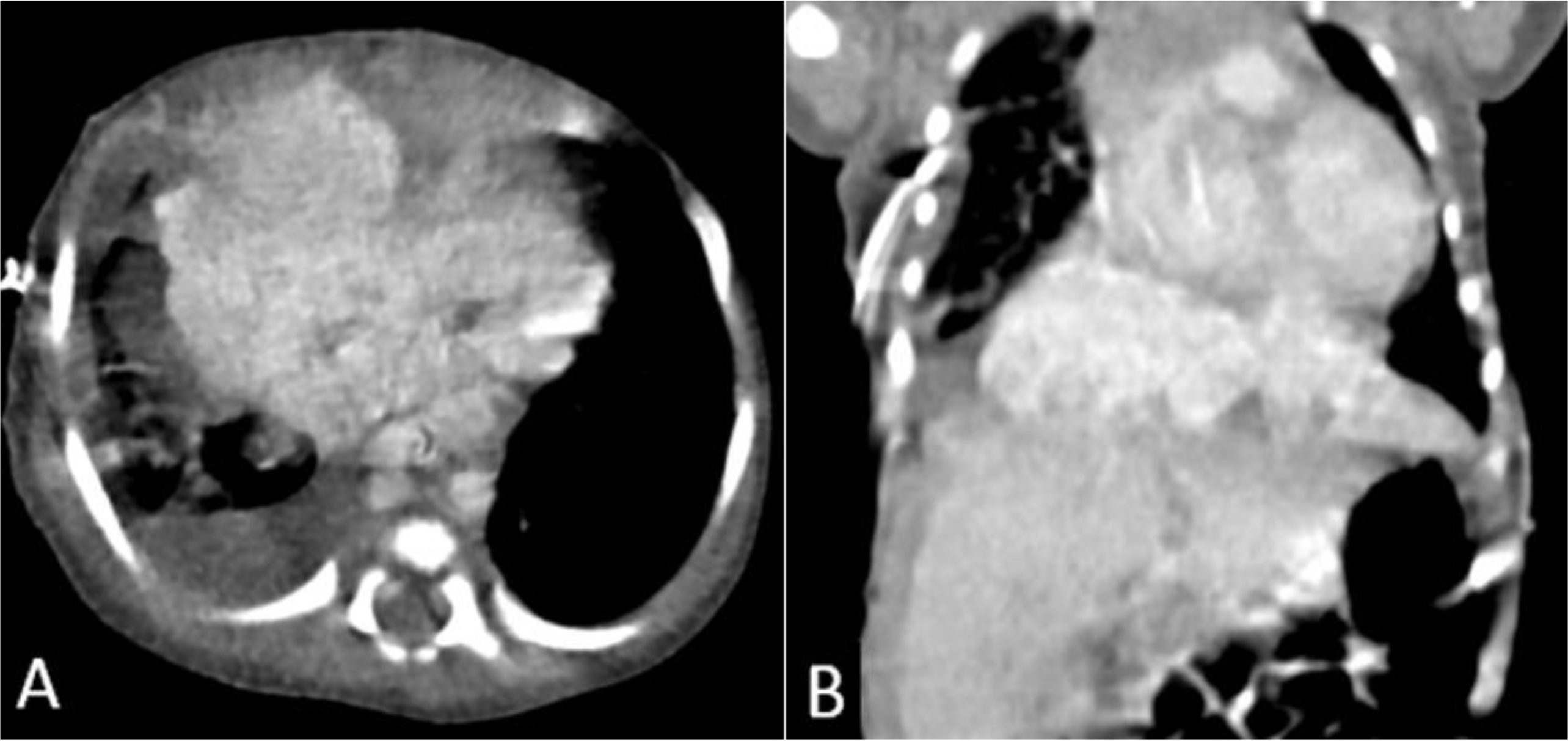
Fig. 3
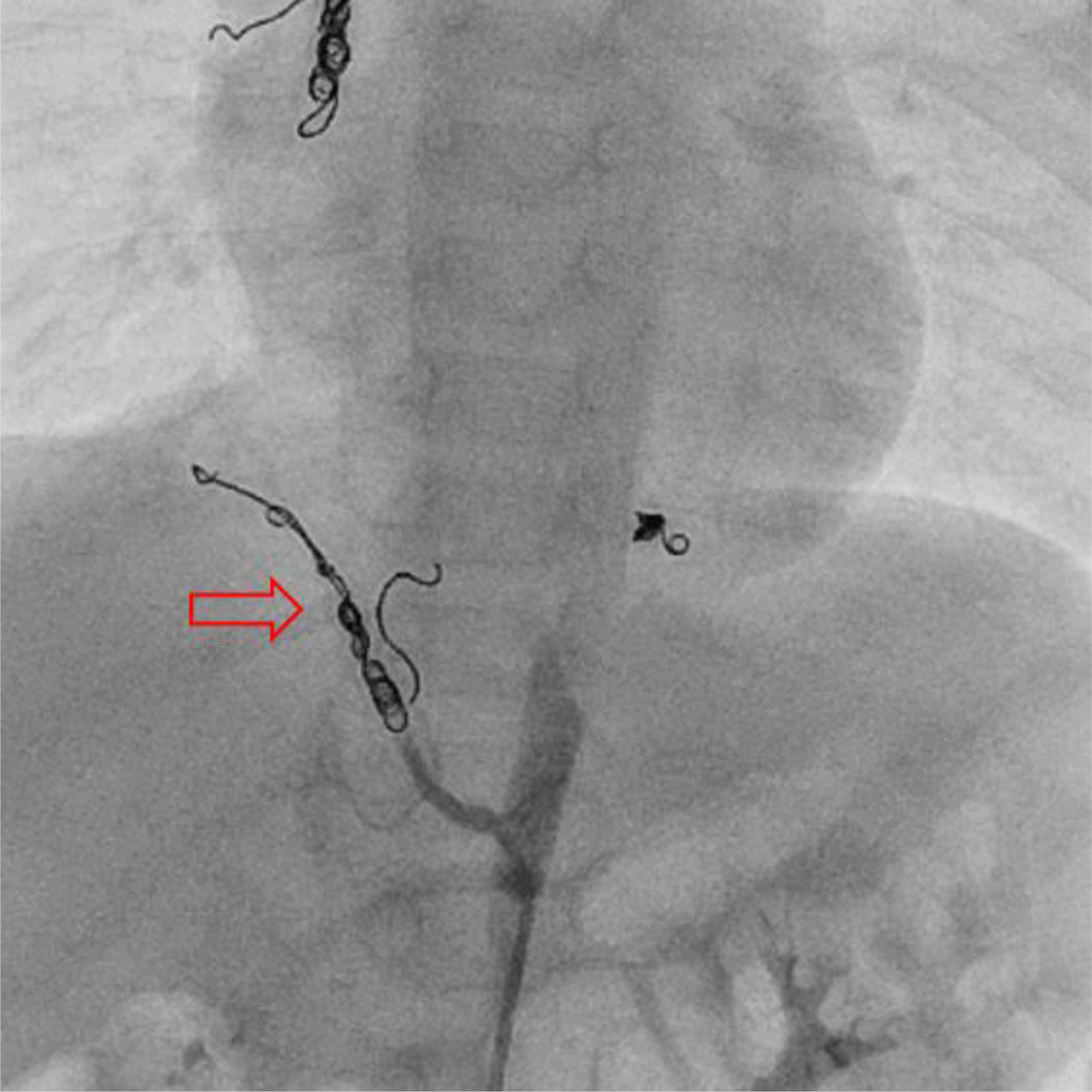
(A) Thoracic aortography showed inferior vena cava interruption and hemagiomatous mass supplied by right internal mammary artery and both inferior phrenic arteries. (B) Coil embolization. Right internal mammary artery was embolized with Interlock coils (4 mm×8 cm, 3 mm×6 cm) (empty arrow). Left inferior phrenic artery was embolized with Tornado coil (3 mm×2 cm).
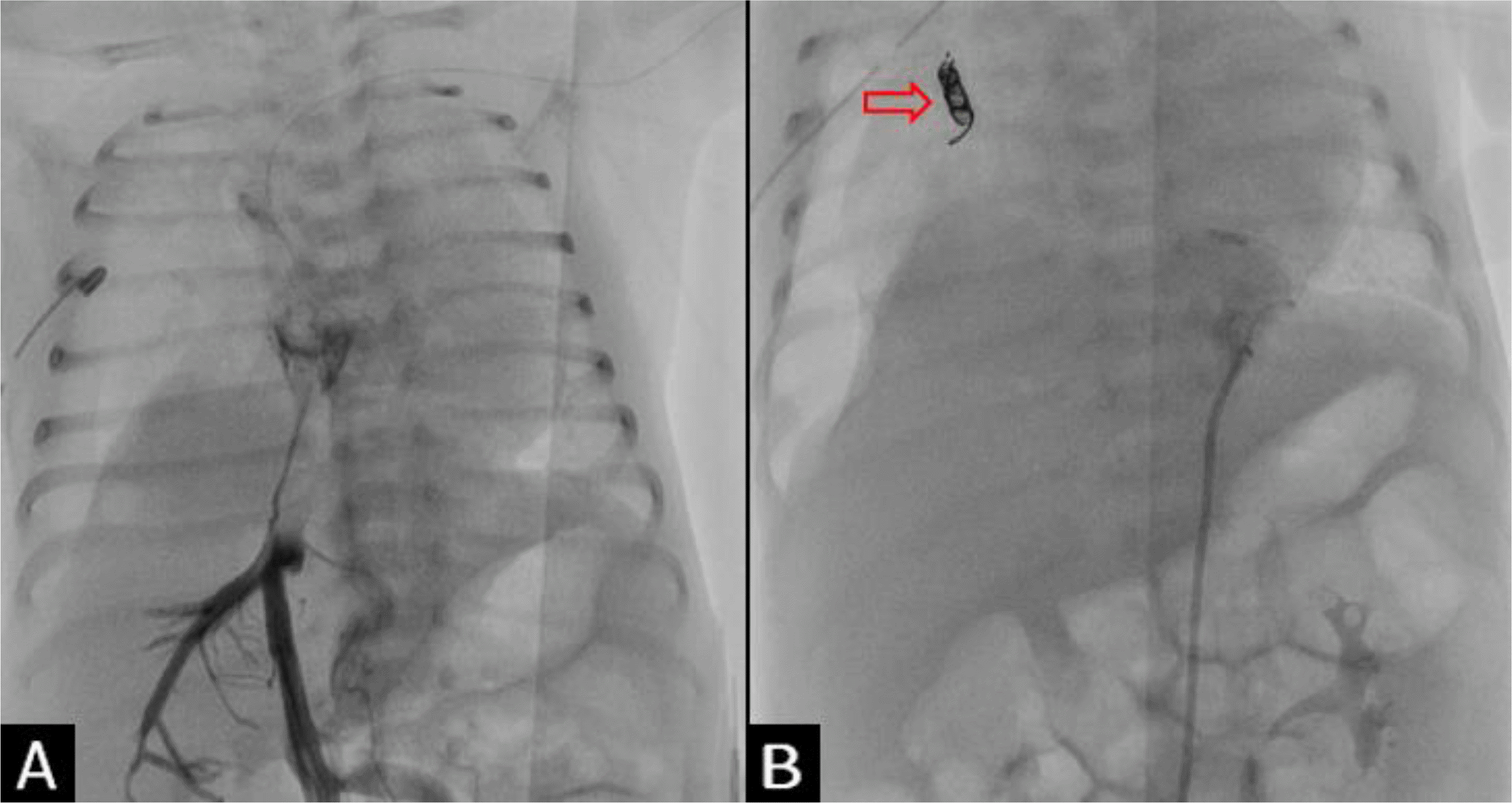
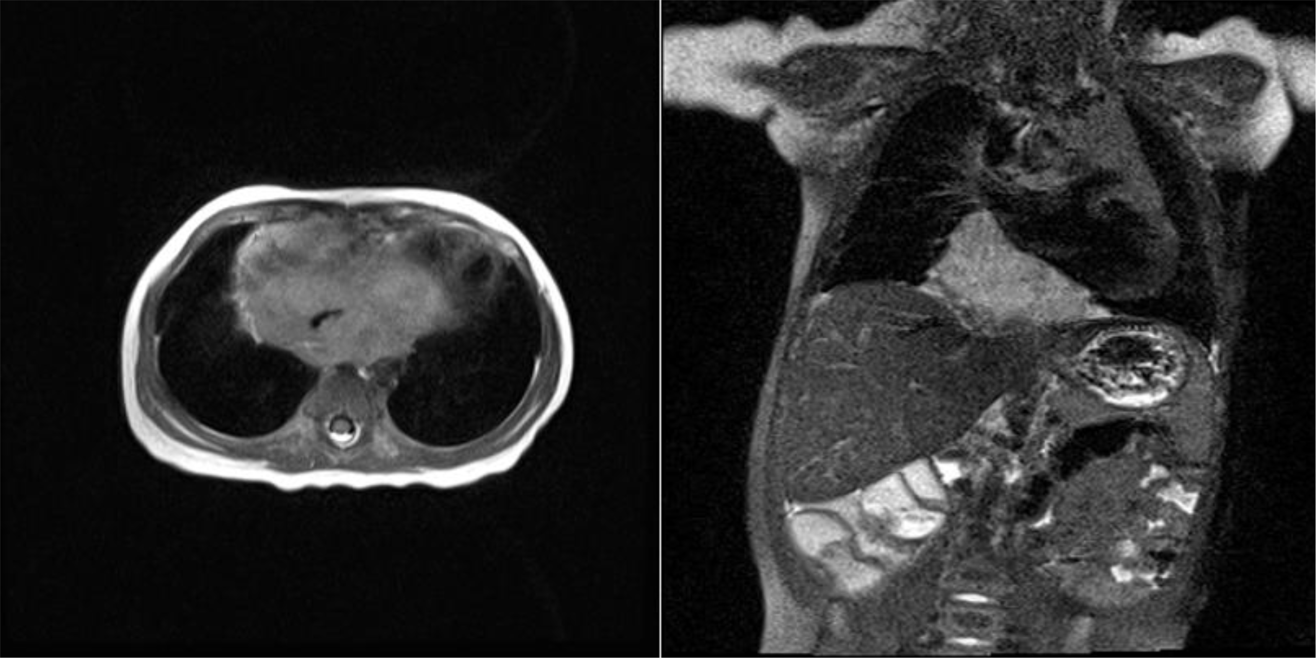
Fig. 4
Multiple purpuric to erythematous nodules and macules over the right cheek, right foot, posterior neck, and left preauricular region.


 PDF
PDF ePub
ePub Citation
 Print
Print


 XML Download