Abstract
Lissencephaly is a cerebral cortical malformation characterized by partial to total loss of gyri and sulci of brain leading to mental retardation and epilepsy. It is caused by failure of division and migration of neural cell in early embryonic period. Microdeletion or point mutation of LIS1 gene is mostly associated with type 1 lissencephaly. A newborn who presented abnormal face and unilateral facial nerve palsy was born and showed near complete agyria on brain MRI. She also has other cranial nerve palsies and ocular abnormalities (aniridia, microcornea and optic disc coloboma). Although we could not found a deletion of LIS1 gene, a case of type 1 lissencephaly with ocular abnormality and cranial nerve palsies is very rare. Therefore we report this case with literature investigations.
REFERENCES
1). Kerjan G., Gleeson JG. Genetic mechanisms underlying abnormal neuronal migration in classical lissencephaly. Trends Genet. 2007. 23:623–30.

2). Cardoso C., Leventer RJ., Matsumoto N., Kuc JA., Ramocki MB., Mewborn SK, et al. The location and type of mutation predict malformation severity in isolated lissencephaly caused by abnormalities within the LIS1 gene. Hum Mol Genet. 2000. 9:3019–28.

3). Hong SE., Shugart YY., Huang DT., Shahwan SA., Grant PE., Hourihane JO, et al. Autosomal recessive lissencephaly with cerebellar hypoplasia is associated with human RELN mutations. Nat Genet. 2000. 26:93–6.

4). Friocourt G., Poirier K., Rakić S., Parnavelas JG., Chelly J. The role of ARX in cortical development. Eur J Neurosci. 2006. 23:869–76.

5). Squier MV. Development of the cortical dysplasia of type II lissencephaly. Neuropathol Appl Neurobiol. 1993. 19:209–13.

6). Santavuori P., Somer H., Sainio K., Rapola J., Kruus S., Nikitin T, et al. Muscleeye-brain disease (MEB). Brain Dev. 1989. 11:147–53.

7). Kimura S., Sasaki Y., Kobayashi T., Ohtsuki N., Tanaka Y., Hara M, et al. Fukuyama-type congenital muscular dystrophy and the Walker-Warburg syndrome. Brain Dev. 1993. 15:182–91.

8). Dobyns WB. The clinical patterns and molecular genetics of lissencephaly and subcortical band heterotopia. Epilepsia. 2010. 51(Suppl 1):5–9.

9). de Rijk-van Andel JF., Arts WFM., Barth PG., Loonen MCB. Diagnostic features and clinical signs of 21 patients with lissencephaly type 1. Dev Med Child Neurol. 1990. 32:707–17.
10). Cho GJ., Oh MJ., Kwon JA., Kim KA., Lee KJ., Hur JY, et al. A case report of Miller-Dieker syndrome. Korean J Perinatol. 2005. 16:181–6.
11). Na JM., Park CH., Yoo EJ., Jung K., Kim KS., Kim YW, et al. A case of MillerDieker syndrome with infantile spasm and Lennox-Gastaut syndrome. J Korean Child Neurol Soc. 2008. 16:86–91.
12). Gleeson JG., Allen KM., Fox JW., Lamperti ED., Berkovic S., Scheffer I, et al. Doublecortin, a brain-specific gene mutated in human X-linked lissencephaly and double cortex syndrome, encodes a putative signaling protein. Cell. 1998. 92:63–72.

13). Shaw MW., Falls HF., Neel JV. Congenital aniridia. Am J Hum Genet. 1960. 12(4 Pt 1):389–415.
14). Hoyt CS., Taylor D. Pediatric ophthalmology and strabismus. 4th ed.London: Saunders Ltd;2013. p. 304–6.
15). Nelson LB., Olitsky SE. Harley's pediatric ophthalmology. 6th ed.Philadelphia: Lippincott Williams & Wilkins;2014. p. 323–5.
16). Gastaut H., Pinsard N., Raybaud CH., Aicardi J., Zifkin B. Lissencephaly (agyria-pachygyria): clinical findings and serial EEG studies. Dev Med Child Neurol. 1987. 29:167–80.
17). Ghai S., Fong KW., Toi A., Chitayat D., Pantazi S., Blaser S. Prenatal US and MR imaging findings of lissencephaly: review of fetal cerebral sulcal development. Radiographics. 2006. 26:389–405.

18). Pilz DT., Dalton A., Long A., Jaspan T., Maltby EL., Quarrell OW. Detecting deletions in the critical region for lissencephaly on 17p13.3 using fluorescent in situ hybridisation and a PCR assay identifying a dinucleotide repeat polymorphism. J Med Genet. 1995. 32:275–8.

Fig. 1
The patient's appearance showing high prominent forehead, hypertelorism and Rt. facial nerve palsy.
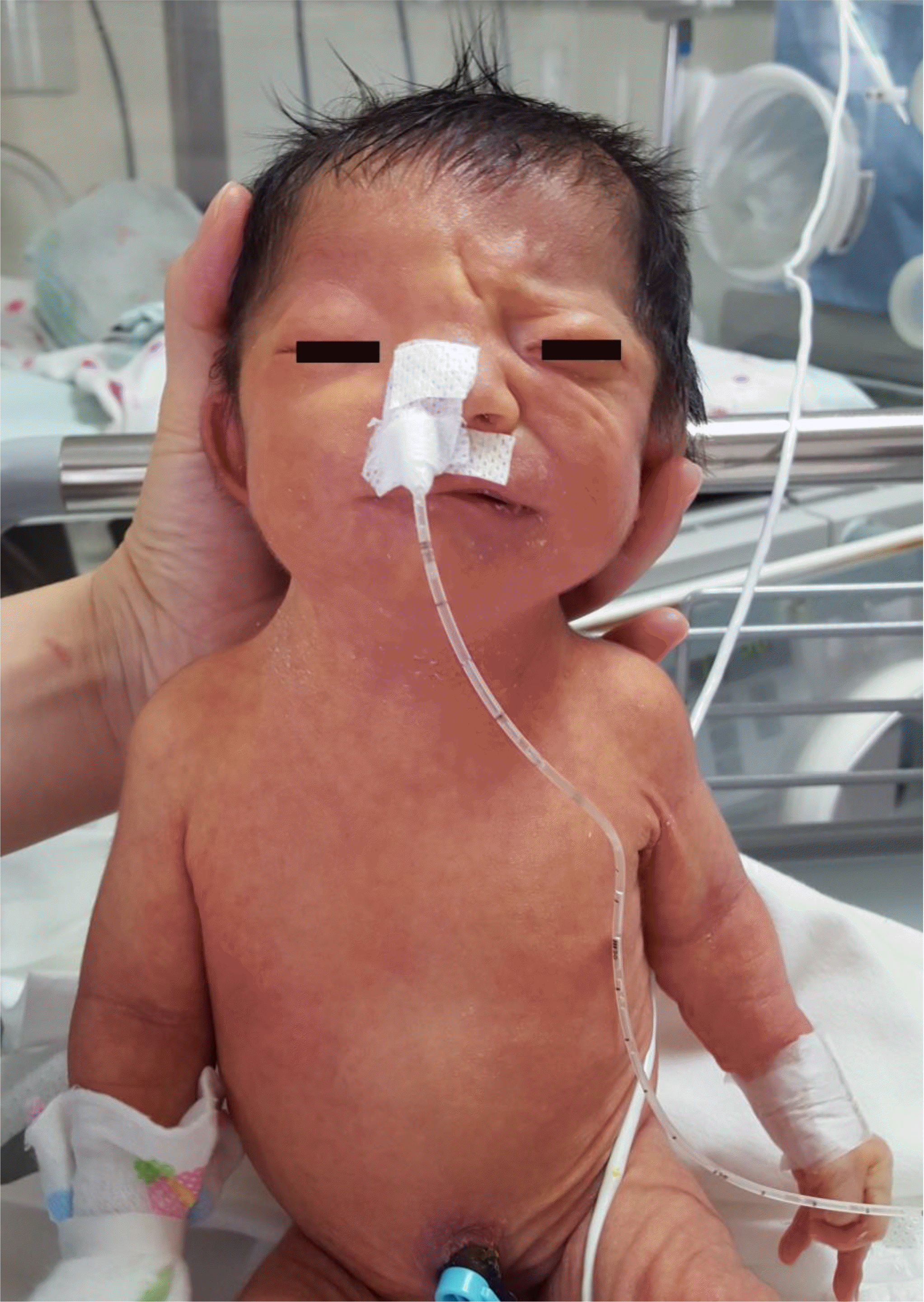
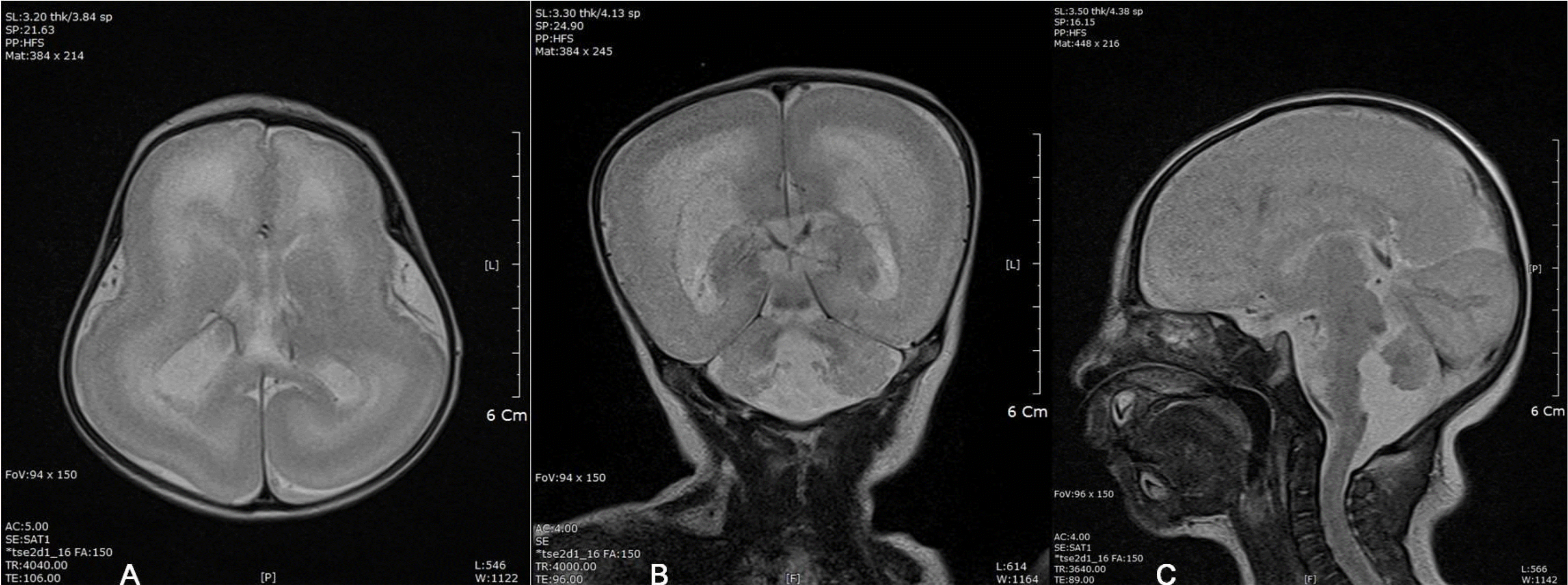
Fig. 3
The patient's brain magnetic resonance imaging T2W axial image (A), coronal image (B), sagittal image (C) showing near complete agyria with doube cortex in both cerebral hemispheres.


 PDF
PDF ePub
ePub Citation
 Print
Print


 XML Download