

 PDF
PDF ePub
ePub Citation
 Print
Print


Abstract
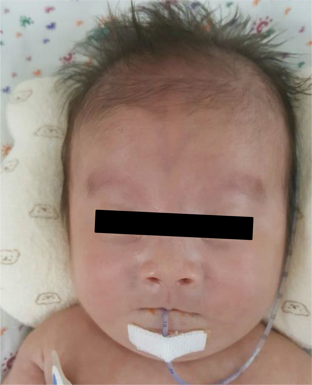
Miller-Dieker syndrome (MDS) is characterized by severe lissencephaly and facial dysmorphism including the prominent forehead, bitemporal hollowing, a short nose with upturned nares, a protuberant upper lip, and a small jaw. MDS can be caused by deletions or mutations of the LIS1 gene on 17p13.3. Our patient was born at 28+3 weeks gestation and weight 950 g at birth. He was suffered from respiratory distress syndrome and feeding intolerance. And he showed colpocephaly with agenesis of corpus callosum on the first brain ultrasonography. So, brain magnetic resonance imaging was performed 2 months of age and revealed agyria, colpocephaly, agenesis of corpus callosum and band heterotopias. At initial physical examination revealed no specific abnormal findings. However, hypotonia and abnormal facial morphology including prominent forehead, a short nose with upturned nares and protuberant upper lip were developed gradually as he got older. Chromosomal microarray was performed and confirmed microdeletion on 17p13.3. In conclusion, MDS should be considered when the baby shows colpocephaly, agenesis of corpus callosum, hypotonia, feeding problem and abnormal facial features even though the baby is preterm infant.
REFERENCES
1). Dobyns WB., Curry CJ., Hoyme HE., Turlington L., Ledbetter DH. Clinical and molecular diagnosis of Miller-Dieker syndrome. Am J Hum Genet. 1991. 48:584–94.
2). Reiner O., Carrozzo R., Shen Y., Wehnert M., Faustinella F., Dobyns WB, et al. Isolation of a Miller-Dieker lissencephaly gene containing G protein beta-subunit-like repeats. Nature. 1993. 364:717–21.
3). Monteagudo A., Timor-Tritsch IE. Development of fetal gyri, sulci and fissures: a transvaginal sonographic study. Ultrasound Obstet Gynecol. 1977. 9:222–8.

4). Miller JQ. Lissencephaly in 2 siblings. Neurology. 1963. 13:841.
5). Garcia CA., Dunn D., Trevor R. The lissencephaly (agyria) syndrome in siblings. Arch Neurol. 1978. 35:608–11.
6). Daube JR., Chou SM. Lissencephaly: two case. Neurology. 1966. 16:179–91.
7). Greenberg F., Courtney KB., Wessels RA., Huhta J., Carpenter RJ., Rich DC, et al. Prenatal diagnosis of deletion 17p13 associated with DiGeorge anomaly. Am J Med Genet. 1988. 31:1–4.
8). Lin CY., Chen CP., Liau CL., Su PH., Tsao TF., Chang TY, et al. Prenatal diagnosis of monosomy 17p (17p13.3→pter) associated with polyhydramnios, intrauterine growth restriction, ventriculomegaly, and Miller-Dieker lissencephaly syndrome in a fetus. Taiwan J Obstet Gynecol. 2009. 48:408–11.
9). McGahan JP., Grix A., Gerscovich EO. Prenatal diagnosis of lissencephaly: Miller-Dieker syndrome. J Clin Ultrasound. 1994. 22:560–3.
10). Chitayat D., Toi A., Babul R., Blaser S., Moola S., Yarkoni D, et al. Omphalocele in Miller-Dieker syndrome: expanding the phenotype. Am J Med Genet. 1997. 69:293–8.
11). Lenzini E., D'Ottavio G., Città A., Benussi DG., Petix V., Peoile V. Perinatal diagnosis of Miller-Dieker syndrome by ultrasound and molecular cytogenetic analysis. Clin Genet. 2007. 72:487–9.
12). Guerrini R., Filippi T. Neuronal migration disorders, genetics, and epilep-togenesis. J Child Neurol. 2005. 20:287–99.
Fig. 1
The initial brain ultrasonography showed colpocephaly and agenesis of corpus callosum at 3 days of age.
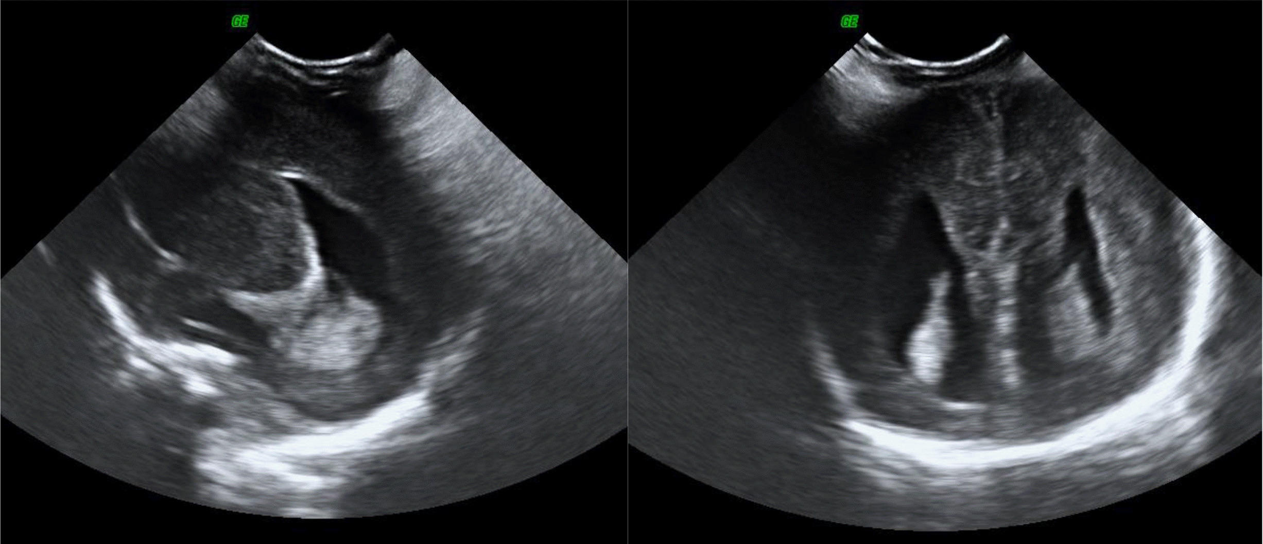
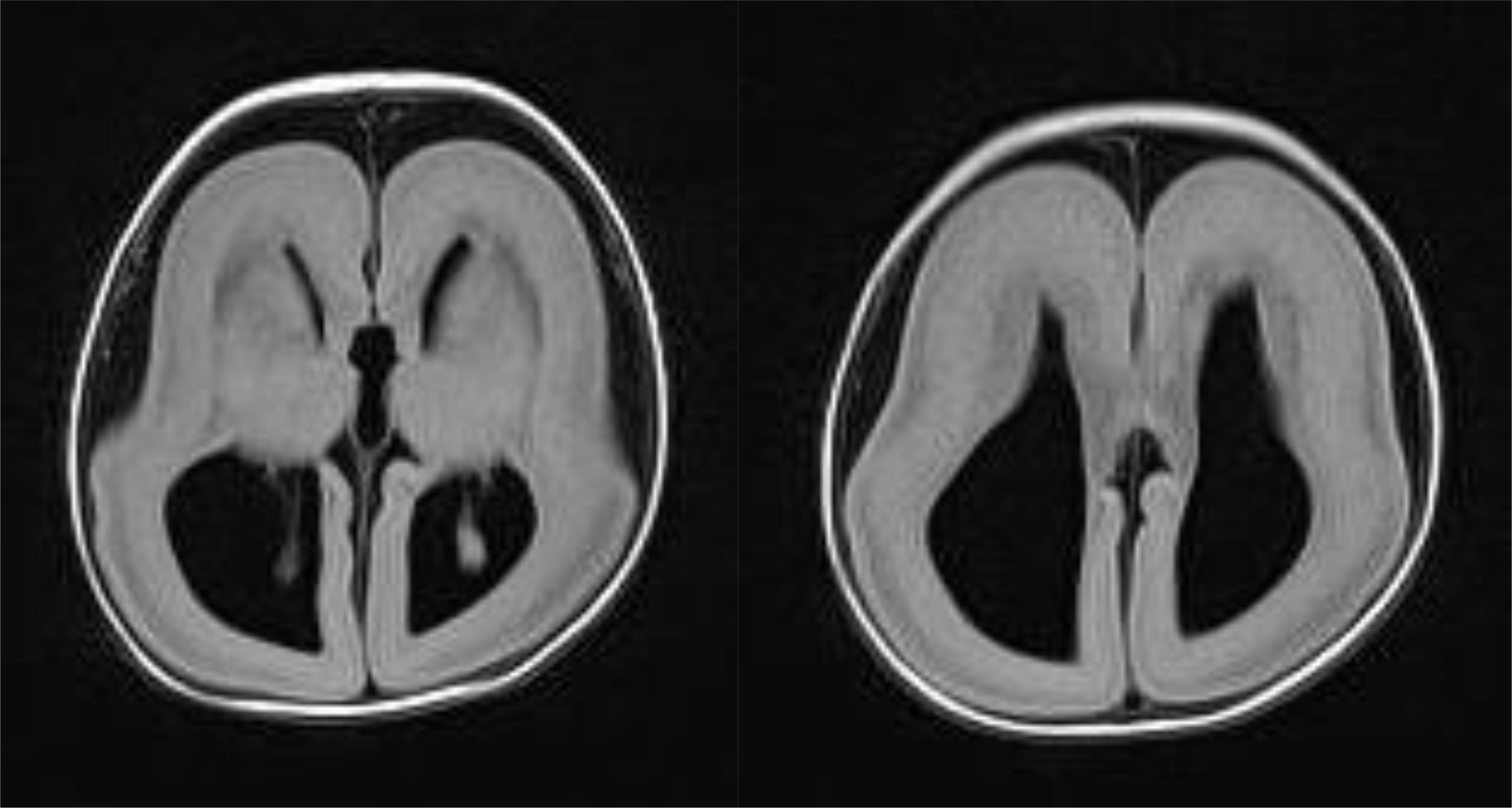
Fig. 2
A brain magnetic resonance imaging showed agyria, colpocephaly, agenesis of corpus callosum and band heterotopia at 2 months of age.
 XML Download