

 PDF
PDF ePub
ePub Citation
 Print
Print


Introduction
In a newborn, the localized collection of air in the mediastinum is caused by congenital mediastinal cystic mass12 or by pneumomediastinum.3 In general, pneumomediastinum occurs in newborns with underlying lung problems, particularly when a ventilator is needed. Pneumomediastinum can also develop spontaneously.4 Congenital mediastinal cystic mass includes congenital cyst, congenital cystic adenomatoid malformation (CCAM) and congenital lobar emphysema (CLE).5 We report a term neonate with spontaneous multiseptated cystic pneumomediastinum suspected as congenital mediastinal cystic mass.
Case report
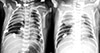
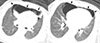
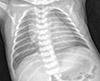
A 3,090 g female neonate was delivered by a spontaneous vaginal delivery at 37 weeks and 4 days gestation, from a 26 year old mother without a specific prenatal history. Her obstetrical history was gravida 2 para 1. The child's Apgar scores were 7 at 1 minute and 8 at 5 minutes after birth. The duration of labor was 6 hours, and the membrane was ruptured just before delivery. Meconium-stained amniotic fluid was not found. The newborn did not need any post-natal resuscitation. After birth, the baby was active, but showed signs of progressive respiratory distress with desaturation, subcostal retractions and nasal flaring. Under an oxygen hood, desaturation improved, but dyspnea continued. The baby was transferred to a neonatal intensive care unit for further evaluation and management. Results of the physical examination were as follows: heart rate 132 beats/min, blood pressure 65/45 mmHg, respiratory rate 64 breaths/min, body temperature 36.5℃ and oxygen saturation 82%. Heart sounds were regular heart beats without murmur. Breathing sounds were not decreased. Subcostal retractions and nasal flaring persisted. Physical examination was otherwise non-specific. Results of the artery blood gas analysis during the inhalation of the oxygen were as follows: pH 7.185, PaCO2 59.8 mmHg, PaO2 99 mmHg and Base deficit -6 mmol/L. Initial complete blood cell counts and, the serum biochemistry profile, including the C-reactive protein, were all within normal limits. The chest X-ray showed a large radiolucent mediastinal lesion on the anterior mediastinum and pneumothorax on both lungs (Fig. 1A). Hence we applied 10 L/min of oxygen via hood. Two hours later, results of the artery blood gas analysis were as follows: pH 7.330, PaCO2 45.1 mmHg, PaO2 62 mmHg and Base deficit -2 mmol/L. Subcostal retractions and nasal flaring decreased. Considering risk of combined congenital infection, empirical antibiotics with ampicillin and gentamicin were used. On the second day of admission, the respiratory rate stabilized to 50 breaths/min. The respiratory distress symptom has decreased visibly. Therefore we stopped the oxygen apply. The oxygen saturation was above 97% without oxygen. On the third day of admission, the baby showed no respiratory distress symptom. Moreover, the baby showed decreased pneumothorax lesions on the chest X-ray. However, the cystic radiolucent lesion on the mediastinum remained (Fig. 1B). A chest CT was performed to determine and characterize the origin of the cystic lesion. The CT revealed multiseptated cystic pneumomediastinum on the anterior mediastinum and over the anterior region of both lungs (Fig. 2). There was no further aggravation in the patient's condition. The patient was discharged at 5 days old. The pneumomediastinum completely disappeared from the chest X-ray taken 2 weeks after discharge (Fig. 3).
Discussion
Pneumomediastinum is defined as the collection of air in the mediastinum. The reported incidence of pneumomediastinum is 4-25 per 10,000 live births.3 This incidence is probably underestimated because many case of spontaneous pneumomediastinum are generally asymptomatic or have minimal symptoms.35
There are two types of pneumomediastinum: a spontaneous type or secondary type related to an underlying lung problem, including assisted ventilation, birth trauma, meconium aspiration, or prematurity.4 The pathogenesis of pneumomediastinum is explained as the increased intra-alveolar pressure causes an alveolar rupture in the perivascular structure and into the mediastinum. When this happens, air dissects the vascular channels and leads to pneumomediastinum.56
In case of pneumomediastinum in neonates, clinical signs are presented as tachypnea, inactivity and, poor oral intake, which are non-specific and unreliable.7 In some cases, heart sounds are decreased.8 Physicians depend on images such as X-rays, chest CT for the accurate diagnosis of pneumomediastinum. Chest radiograph is one of the most frequently performed exams, and one of the essential to diagnose pneumomediastinum.9 Spinnaker Sail Sign is seen on neonatal postero-anterior chest radiograph when thymic lobes are displaced laterally by air resembling a spinnaker sail, it's also called angel wing sign. It's a very typical sign of pneumomediastinum in neonatal age.9 The presence of extra- luminal air within the mediastinum has some radiographic features, namely contour outline of anatomic structures by air seen as lucent lines. Tubular artery sign, continuous diaphragm sign, naclerios's V sign and ring around artery sign are radiographic features of pneumomediastinum.101112 Most cases of pneumomediastinum show spinnaker sail signs that appear as a crescentic figuration of the thymus in chest X- ray.9 However, the chest anteroposterior view findings in our patient did not exhibit the spinnaker sail signs and remained cystic and bubbly lesion on the mediastinum. So we doubt other diseases representing cystic radiolucent lesion on the mediastinum.
The differential diagnosis of the cystic radiolucent lesion on the mediastinum includes pneumomediastinum, congenital mediastinal cyst, CCAM, and CLE.5 More data on the radiolucent cystic area can be obtained by chest CT.13 On chest CT, radiologic feature of pneumomediastinum is small amounts of air appear as linear or curvilinear lucencies outlining mediastinal contours such as subcutaneous emphysema, air anterior to pericardium, air around pulmonary artery, air outlining major aortic branches and bronchial wall.14 In our case, CT revealed multiseptated cystic pneumomediastinum. Our case shows different features compared with typical neonatal pneumomediastinum: tendency to loculate locally and presence of multiple internal septa. The study of Quattromani et al.15 has reported multiloculated multiseptated pneumomediastinum that is similar to our case. They explained the reason of tendency to loculate and multiple internal septa as follows. Mediastinum is restricted by fascial band which covers the thymus and merges with fibrous pericardium. The air dissects within the interlobular and connective tissue septa of the thymic capsule, giving rise to a multiseptated cystic appearance.15
One study has suggested that a surgical approach of pneumomediastinum is necessary in case of progressive cardio-pulmonary distress and failure of supportive treatments.16 However, pneumomediastinum is generally self- limited.16 In our study, only close observation and supportive management are required for the treatment of pneumomediastinum in newborns.
In conclusion, the cystic and bubbly lesion on the mediastinum in chest X-ray suggest congenital mediastinal cystic mass or pneumomediastinum. We could have differential diagnosed spontaneous multiseptated cystic pneumomediastinum from congenital mediastinal cystic mass by using chest CT.
 XML Download