

 PDF
PDF ePub
ePub Citation
 Print
Print


INTRODUCTION
Rheumatoid nodules occur in up to 30 percent of adult patients with rheumatoid arthritis (1). Rheumatoid nodules are frequently found at the level of subcutaneous soft tissue, over prominent bones that are usually exposed to repetitive mechanical irritation, such as the extensor surface of the forearm, fingers, occiput, heel, and elbow (1234). The MRI scan features of rheumatoid nodules are variable, and nodules can be predominantly solid, or contain a cystic portion (23).
Our patient was referred to the hospital, because of buttock swelling and discomfort in seating position, and diagnosed with ischiogluteal bursitis, because of its characteristic location, and a cystic appearance in the MRI scan. The diagnosis of the lesion was confirmed as a rheumatoid nodule histologically.
To the best of our knowledge, no previous reports have described a rheumatoid nodule deep in the muscle, just beneath the bone, as a cystic lesion. We describe in our report, a case of rheumatoid nodule, with cystic change over ischial tuberosity, mimicking ischiogluteal bursitis.
CASE REPORT
A 68-year-old woman with a seven-year history of rheumatoid arthritis (RA) was referred to our institution suffering for six months, from right buttock swelling and discomfort in the seating position.
The patient was presented with symmetrical inflammatory polyarthritis, involving bilateral ankle, elbow, hip, tarsometatarsal, and proximal interphalangeal joints, a condition she has been suffering from for seven years. Serologic test revealed low-positive rheumatoid factor (14.5 IU/mL), and negative anti-cyclic citrullinated peptides (anti-CCP) antibody. Laboratory studies revealed abnormal C-reactive protein (CRP) (27.5 mg/dL), and erythrocyte sedimentation rate (ESR) (112 mm/hr). According to the 2010 ACR/EULAR (American College of Rheumatology/European League Against Rheumatism) criteria for rheumatoid arthritis, the score was 7/10, so the patient was classified as having rheumatoid arthritis.
The patient complained about right buttock swelling, and discomfort on seating position, but had no relevant history of trauma. Needle aspiration was performed at a previous hospital, and aspiration yielded 100 mL of fluid. Regardless of aspiration, the lesion had enlarged again, when the patient was admitted to our institution.
Physical examination revealed a fluctuating mass, measuring 5 × 4 × 4 cm in the right buttock, near the ischial tuberosity. The mass was soft, and manifested mild tenderness. The patient had other cystic lesions on the coccyx and left elbow, which was exposed to repetitive mechanical irritation. The lesion of the left elbow was excised, and histologically diagnosed as a rheumatoid nodule.
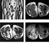
Initial radiographs of the hip and pelvis, demonstrated normal bone alignment, without any osseous abnormality. Subsequently an MRI scan, on a 1.5 Tesla Achieva (Philips, Einthoven, the Netherlands), was performed. The MRI protocol included T1-weighted image (repetition time [TR]/echo time [TE], 1019.6-1165.2/18.0) and T2-weighted image (TR/TE, 5619.5-6426.3/100.0). T2 spectral presaturation with inversion recovery (SPIR) images (TR/TE, 2000.0/70.0), modified proton density images (TR/TE, 4000.0/33.0) and gadolinium enhanced T1-weighted images (TR/TE 1524.5–1742.3/18.0), were included. The studies were performed with a 350 × 350 field-of view, and a 4–6 mm slice thickness, with a 0.9 mm gap. An 8.5 × 8.0 × 5.2 cm well-delineated cystic lesion was noted, extending beneath the gluteus maximus, and located posteroinferior to the ischial tuberosity (Fig. 1a). The lesion was encapsulated with a thick low signal intensity capsule, on T1-weighted and T2-weighted images (Fig. 1b). The lesion revealed heterogeneous high signal intensity on T2-weighted images, suggesting most contents were fluid collection (Fig. 1c). Contrast-enhanced T1-weighted images revealed rim-like peripheral capsular enhancement of the lesion (Fig. 1d). Another cystic lesion was noted on the subcutaneous tissue of left buttock, which was exposed to the mechanical pressure (Fig. 1). Imaging findings of the lesion were similar to those of the lesion of ischial tuberosity. The subcutaneous cystic lesion on left buttock can be diagnosed as a rheumatoid nodule with central necrosis, considering imaging findings and the patient's history. Radiologic diagnosis of the lesion abutting to the ischial tuberosity was noted as chronic complicated ischiogluteal bursitis, and the patient underwent excision of the mass.
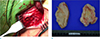
During surgery, the lesion was observed extending from the subcutaneous tissue, to the deep portion of muscle, abutting on the posteroinferior portion of ischial tuberosity (Fig. 2a). The mass was surrounded by a firm and thick capsule, containing yellowish fluid, grossly (Fig. 2b).
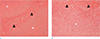
Histopathologically, the inner side of the cyst revealed a large amount of fibrinoid necrotic material surrounded by histiocytes and fibrosis (Fig. 3). Neither lining cells, nor extensive lymphoplasmacytic reaction, is noted. The lesion extended to the dermis. Several foci of nodular granuloma-like lesion with central necrosis were noted in the subcutis. Mycobacterial staining and polymerase chain reaction were negative. Histological analysis revealed the lesion, consistent with the rheumatoid nodule similar to that found subcutaneously.
DISCUSSION
The skin is the most common extra-articular target of rheumatoid arthritis (1). Rheumatoid nodules are frequently localized at sites of external pressure or repetitive irritation, such as the extensor surfaces of the hand, heel, sacral prominences, ears, and nose (15). To the best of our knowledge, there has been only one case report of a rheumatoid nodule, located in the skeletal muscle close to the fascia (6). However, there have been no reports of rheumatoid nodules extending deep into the muscle, and adjacent to the bone.
In our case, the nodule was cystic and extended deep into the gluteus maximus muscle, abutting on the surface of the ischial tuberosity, typical for ischiogluteal bursitis (7). However, histopathologic finding of the case, was composed of fibrinoid degeneration of collagen, surrounded by histiocytes and giant cell reaction, consistent with a rheumatoid nodule.
On pathology, rheumatoid nodule is a subcutaneous lesion, revealing granuloma-like reaction with central necrosis. Old rheumatoid nodule may show cystic degeneration of the necrotic component that has progressed beyond the “fibrinoid stage” and looks like a cystic nodule (235).
The cystic form of rheumatoid nodule is composed of three zones; 1) a central necrotic zone; 2) an intermediate zone of radially arranged elongated cells, with pale nuclei and indistinct outlines; and 3) an outer zone of chronic inflammatory cells, in which plasma cells are numerous (235).
Synovitis in RA also affects the synovial lining of the bursa, which results in bursitis (8). Characteristic histopathological features of RA synovitis are summarized as follows: 1) moderate to marked proliferation of synovial lining layer, often accompanied by a typical palisading of synovial cells; 2) presence of non-foreign body type multinucleated giant cells, including Langhans giant cells; 3) diffuse infiltration of lymphocytes and plasma cells, and the frequent formation of lymphoid follicles and a germinal center; 4) marked proliferation of blood vessels; and 5) hemosiderosis in the synovial stroma and synovial cells (9).
Histopathology relative to our case report, revealed fibrocollagenous tissue infiltrated with necrotizing granuloma, with central fibrinoid necrosis, which was compatible to the histology of the rheumatoid nodule with a cystic component. There was an absence of proliferation of synovial cells, and diffuse infiltration of lymphocytes and plasma cells with formation of lymphoid follicles, which were characteristic features of RA synovitis.
The MRI scan features of rheumatoid nodules correlate with the underlying histopathological evolution of these lesions. The nodule can be seen as a predominantly solid nodule, or a nodule that contains a cystic portion (23).
The nodule was hypointense in T1- and T2-weighted images and hyperintense in contrast-enhanced images. Histologically, this type of lesion is composed entirely of chronic inflammatory cells such as histiocytes, macrophages, and fibroblasts, and small vessels.
The other type of nodule with central necrosis, can be noted as heterogeneously hyperintense in T2-weighted images. The cystic components are surrounded by a rim of solid nodular tissue that is hypointense in T1- and T2-weighted images. On contrast-enhanced scan, a rim of enhancing tissue containing inflammatory infiltrates is clearly demarcated from the non-enhancing hypointense necrotic tissue. The microscopic sections from the nodule revealed a centrally necrotic cystic area, surrounded by palisading inflammatory cellular infiltrate, containing predominantly macropahges, lymphocytes, and plasma cells, in addition to fibroblasts and small vessel (23).
The MRI findings of ischiogluteal bursitis may be nonspecific. The lesion reveals hyperintense in T2-weighted images, hypointense in T1-weighted images, and peripheral rim enhancement on contrast-enhanced scan (7). The bursitis may reveal variable signal intensity in T1- and T2-weighted images, according to the content of the bursal sac. The typical location of ischiogluteal bursitis might be a very important diagnostic criterion, for separating bursitis from the other cystic masses. It is typically located deep in the inferior portion of the gluteus maximus muscle, and posterior inferiorly, to the ischial tuberosity, on the sagittal sections. The superior end of the bursa abuts to the inferomedial surface of the ischial tuberosity (7).
In our case report, the lesion revealed a cystic lesion, a nonspecific finding, which was observed in the bursitis and rheumatoid nodules with complete central necrosis (237). The typical location of the cystic lesion, deep into the gluteus maximus muscle and abutting the ischial tuberosity, implies ischiogluteal bursitis, rather than a rheumatoid nodule. However, the following are diagnostically helpful findings that suggest rheumatoid nodule rather than ischiogluteal bursitis: 1) the patient's history of RA; 2) other same-appearing lesions on coccyx, buttock, and elbow, one of which was confirmed as rheumatoid nodule; and 3) the extent of the lesion not confined to the level abutting to the bone, but extending to the subcutaneous layer.
In conclusion, a rheumatoid nodule located not only at the subcutaneous tissue, but extending to the deep portion of muscle adjacent to the bone, is rare. Awareness of the imaging findings and rare location will be valuable in correct diagnosis and treatment.
 XML Download