Abstract
South Korea has the highest reported suicide rate among all countries belonging to the Organization for Economic Cooperation and Development. Nitrogen is a colorless, odorless and nontoxic gas. Nitrogen gas has, however, been recently used as a method of attempted suicide, its nontoxity notwithstanding. We herein report on an unusual case involving a 30-year-old male who presented with symptoms after a suicide attempt by nitrogen inhalation. Diffusion-weighted imaging of his brain was showed curvilinear high signal intensity in the bilateral frontal and right occipital cortices, with subtle low apparent diffusion coefficient value. In addition, T2-weighted images and fluid attenuated inversion recovery images revealed subtle high signal intensity in the bilateral frontal cortices, basal ganglia and occipital cortices with contrast enhancement.
According to the Organization for Economic Cooperation and Development (OECD) report in 2015, South Korea had the highest suicide rate among all countries that belong to the OECD.
In contrast to the pattern in most OECD countries, death rates from suicide in Korea have risen significantly in the last decade (1). Recently, several organizations and internet communities in favor of assisted suicide have promoted the use of nitrogen (N2) gas to that end (2). Nitrogen gas has caused accidental deaths in industrial or laboratory explosion, and during scuba diving and anesthesia (2). Although it is reported that industrial nitrogen asphyxiation hazards resulted in 80 deaths during the period 1992 through 2002, there is a paucity of documentation regarding nitrogen gas as a means of committing suicide (23). Nitrogen is a colorless, odorless, nontoxic, and generally inert gas that is a normal component (78.09%) of the atmosphere, at standard temperature and pressure (4). However, nitrogen can be hazardous when it displaces oxygen resulting in hypoxic damage (23). Nitrogen intoxication manifests with various symptoms such as progressive fatigue, loss of coordination, purposeful movement and balance, nausea, a complete inability to move and unconsciousness (24). Here, we describe a case of brain magnetic resonance imaging (MRI) findings associated with nitrogen gas inhalation, which have been rarely reported previously.
A 30-year-old man visited the emergency department with complaint of numbness of the bilateral upper extremities. He had a past medical history of a diagnosed “gambling disorder”. He reported that a week before, he attempted suicide by inhaling pure nitrogen gas with people he had met through an internet suicidal community, however, he did not present with any symptoms. His stated reason for attempting suicide was financial difficulty. He reported that five days prior to the emergency room visit, he attempted suicide again, on this occasion by inhaling nitrogen gas through a plastic bag. And after that he lost consciousness for a while. A few hours later, he recovered consciousness but awoke with symptoms of diplopia, headache and stiffness of both hands with slow progression over the course of the past three days. On hospital visiting day, he presented with complaint of numbness and cramping of both hands.
His vital signs were stable on admission. His laboratory tests were all within normal range including the hemoglobin level (13.8 g/dL), partial pressure of oxygen in the arterial blood (PaO2) (107 mmHg), and saturation of oxygen in the arterial blood (SaO2) (98.4%).
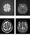
An initial brain computed tomography (CT) was obtained and revealed no significant abnormality. Diffusion-weighted imaging (DWI) of the brain (Magnetom Avanto 1.5T, Siemens, Erlangen, Germany) was obtained and showed curvilinear high signal intensity in the bilateral frontal and right occipital cortices with subtle low apparent diffusion coefficient (ADC) value (Fig. 1).
On hospital day five, electroencephalography was performed and showed no abnormality.
MRI was obtained on a 3.0T system (Achieva, Philips Healthcare, Best, The Netherlands) on hospital day ten. T2-weighted images (T2WI) and fluid attenuated inversion recovery (FLAIR) images revealed subtle high signal intensity in the bilateral frontal cortices, basal ganglia (Fig. 2) and occipital cortices (Fig. 3). The lesions of the occipital cortex show irregular enhancement on the contrast-enhanced T1-weighted images (T1WI) (Fig. 3).
The patient's symptoms improved with supportive care and psychiatric management. He was discharged, without any documented neurological deficits, on hospital day fifteen.
Suicide has become a critical issue in South Korea, according to the OECD report (1). Potential means and methods of suicide commonly appear on web searches and are easily accessed over the internet (5). Nitrogen gas as a means of suicide was invented by Dr. Philip Nitschke in 2007, and has been frequently and widely described since that time (2). Last year, suicide by nitrogen gas received coverage on the news in Korea. Nitrogen is safe to breathe only when mixed with the appropriate amount of oxygen. Nitrogen is a colorless, odorless, nontoxic and generally inert gas that is a normal component (78.09%) of the atmosphere, at standard temperature and pressure (4). However commercial nitrogen gas is usually stored in large cylinders (2). These pure nitrogen gas can be hazardous when it replaces oxygen and causes various symptoms such as progressive fatigue, nausea, partial or complete physical paralysis and/or unconsciousness (2, 3). Nitrogen gas has caused accidental deaths in industrial settings and laboratory explosions, as well as during scuba diving and surgical anesthesia (2). When a diver rapidly ascends from depth, nitrogen gas bubbles form in the tissues and bloodstream (nitrogen narcosis). Nitrogen gas embolisms usually present, radiographically and clinically, with a stroke-like appearance of the gray matter, and can also cause white matter abnormalities due to the high lipidsolubility of nitrogen (6). There are, however, few if any radiographic reports reflecting MRI findings arising, purely and solely, from nitrogen gas inhalation. Furthermore, the incident of nitrogen gas inhalation related to the suicidal attempt was reported from a medical-legal perspective. These reports have described only autopsy - postmortem - findings and there are very few reports regarding survivors of nitrogen gas inhalation in the standard atmosphere (24). The hypoxia triggered by pure nitrogen inhalation is associated with serious complications affecting the brain, and it is critical to recognize the imaging findings which are specific to nitrogen intoxication (7).
In our case, DWI and FLAIR high signal intensity lesions were observed in the brain cortex. These MRI findings are identical when compared with those produced by the hypoxic injury. In moderate-to-severe cases of hypoxic encephalopathy, vulnerable areas are the brain cortex, especially the perirolandic, and medial occipital cortices with precentral gyri, and these findings are probably due to cytotoxic edema (78). Cortical enhancement is usually seen after a few weeks, and is likely due to breakdown of the blood-brain barrier and impaired autoregulation. However, it is thought that early gyral contrast enhancement could be related to the severity, or extent, of the hypoxic brain damage leading to the breakdown of the blood brain barrier and reperfusion of the hypoxic ischemic brain (9).
Tur et al. (10) reported on a case of nitrogen gas inhalation which occurred in the context of an industrial accident. It was noted that the patient initially presented with altered mental status and involuntary movement. After high-flow oxygen therapy, the patient was awake and alert ten hours after the incident, and was eventually discharged without residual neurologic deficit. It is similar to our patient's clinical presentation. Treatment of nitrogen intoxication mainly consists of supportive care and a concerted effort to prevent or obviate any additional or ongoing injury (8). In addition, nitrogen gas is lighter than air. Therefore, it disperses quickly in the atmosphere. Therefore and although nitrogen gas may serve as an effective means of committing suicide, this method of selfmurder would not prove inimical to the health of, or fatal, to anyone that might happen to stand next to the body during recovery (2). Furthermore, any patient who has attempted suicide, should receive appropriate psychiatric intervention and treatment (5).
Other gases used in suffocation and suicide are more commonly-encountered gases such as carbon dioxide, carbon monoxide and methane that result in depression of the central nervous system by exclusion of oxygen (45). In some cases, the method of the attempted suicide is difficult to determine as often, would-be suicide victims arrive in a state of unconsciousness or if conscious, they are embarrassed or otherwise unwilling to provide a complete or truthful medical history or explanation for their current condition. However, some gases do produce specific and characteristic imaging findings on brain MRI. Carbon monoxide most often involves the globus pallidus, although the cerebral white matter and basal ganglia are frequently involved as well (5). If brain MRI findings of carbon monoxide inhalation involve other basal ganglia, it is difficult to make a differential diagnosis, from possible nitrogen inhalation or other deep anoxic injury. It has been determined that the caudate and putamen are the most vulnerable in hypoxic insult (57).
In conclusion, we are reporting on a rare case of nitrogen inhalation occasioned by a failed suicide attempt which, on radiographic examination, presented as DWI and FLAIR high signal intensity in the frontal and occipital cortices with contrast enhancement of occipital cortices. Awareness and sensitivity to these attributes, these specific characteristics, will hopefully allow for earlier diagnosis and optimal management of the sequelae of acute nitrogen inhalation brain injury.
Figures and Tables
Fig. 1
A 30-year-old man after suicide attempt by nitrogen inhalation through a plastic bag. (a,b) Diffusion-weighted image shows curvilinear high signal intensity (SI) in the bilateral frontal and right occipital cortices. (c, d) Apparent diffusion coefficient map shows low value in the bilateral frontal and right occipital cortices.

References
1. OECD. Health at a Glance 2015: OECD indicators. Paris: OECD Publishing;2015.
2. Madentzoglou MS, Kastanaki AE, Nathena D, Kranioti EF, Michalodimitrakis M. Nitrogen-plastic bag suicide: a case report. Am J Forensic Med Pathol. 2013; 34:311–314.
3. USCSB. Safety Bulletin: Hazards of Nitrogen Asphyxiation, No. 2003-10-B. June 2003; 2003.
4. Harding BE, Wolf BC. Case report of suicide by inhalation of nitrogen gas. Am J Forensic Med Pathol. 2008; 29:235–237.

5. DiPoce J, Guelfguat M, DiPoce J. Radiologic findings in cases of attempted suicide and other self-injurious behavior. Radiographics. 2012; 32:2005–2024.

6. Kamtchum Tatuene J, Pignel R, Pollak P, Lovblad KO, Kleinschmidt A, Vargas MI. Neuroimaging of diving-related decompression illness: current knowledge and perspectives. AJNR Am J Neuroradiol. 2014; 35:2039–2044.

7. White ML, Zhang Y, Helvey JT, Omojola MF. Anatomical patterns and correlated MRI findings of non-perinatal hypoxic-ischaemic encephalopathy. Br J Radiol. 2013; 86:20120464.

8. Huang BY, Castillo M. Hypoxic-ischemic brain injury: imaging findings from birth to adulthood. Radiographics. 2008; 28:417–439.

9. Maurya VK, Ravikumar R, Bhatia M, Rai R. Hypoxicischemic brain injury in an adult: magnetic resonance imaging findings. Med J Armed Forces India. 2016; 72:75–77.

10. Tur FC, Aksay E. Asphyxia due to accidental nitrogen gas inhalation: a case report. Hong Kong J Emerg Med. 2012; 19:46–48.


 PDF
PDF ePub
ePub Citation
 Print
Print


 XML Download