

 PDF
PDF ePub
ePub Citation
 Print
Print


Abstract
Purpose
To report the use of multiband accelerated echo-planar imaging (EPI) for resting-state functional MRI (rs-fMRI) to achieve rapid high temporal resolution at 3T compared to conventional EPI.
Materials and Methods
rs-fMRI data were acquired from 20 healthy right-handed volunteers by using three methods: conventional single-band gradient-echo EPI acquisition (Data 1), multiband gradient-echo EPI acquisition with 240 volumes (Data 2) and 480 volumes (Data 3). Temporal signal-to-noise ratio (tSNR) maps were obtained by dividing the mean of the time course of each voxel by its temporal standard deviation. The resting-state sensorimotor network (SMN) and default mode network (DMN) were estimated using independent component analysis (ICA) and a seed-based method. One-way analysis of variance (ANOVA) was performed between the tSNR map, SMN, and DMN from the three data sets for between-group analysis. P < 0.05 with a family-wise error (FWE) correction for multiple comparisons was considered statistically significant.
Results
One-way ANOVA and post-hoc two-sample t-tests showed that the tSNR was higher in Data 1 than Data 2 and 3 in white matter structures such as the striatum and medial and superior longitudinal fasciculus. One-way ANOVA revealed no differences in SMN or DMN across the three data sets.
Conclusion
Within the adapted metrics estimated under specific imaging conditions employed in this study, multiband accelerated EPI, which substantially reduced scan times, provides the same quality image of functional connectivity as rs-fMRI by using conventional EPI at 3T. Under employed imaging conditions, this technique shows strong potential for clinical acceptance and translation of rs-fMRI protocols with potential advantages in spatial and/or temporal resolution. However, further study is warranted to evaluate whether the current findings can be generalized in diverse settings.
References
1. Moeller S, Yacoub E, Olman CA, et al. Multiband multislice GE-EPI at 7 tesla, with 16-fold acceleration using partial parallel imaging with application to high spatial and temporal whole-brain fMRI. Magn Reson Med. 2010; 63:1144–1153.

2. Setsompop K, Gagoski BA, Polimeni JR, Witzel T, Wedeen VJ, Wald LL. Blipped-controlled aliasing in parallel imaging for simultaneous multislice echo planar imaging with reduced g-factor penalty. Magn Reson Med. 2012; 67:1210–1224.
3. Smith SM, Miller KL, Moeller S, et al. Temporally-independent functional modes of spontaneous brain activity. Proc Natl Acad Sci U S A. 2012; 109:3131–3136.
4. Cole DM, Smith SM, Beckmann CF. Advances and pitfalls in the analysis and interpretation of resting-state FMRI data. Front Syst Neurosci. 2010; 4:8.
5. Cordes D, Haughton VM, Arfanakis K, et al. Frequencies contributing to functional connectivity in the cerebral cortex in "resting-state" data. AJNR Am J Neuroradiol. 2001; 22:1326–1333.
6. Leopold DA, Murayama Y, Logothetis NK. Very slow activity fluctuations in monkey visual cortex: implications for functional brain imaging. Cereb Cortex. 2003; 13:422–433.
7. Malinen S, Vartiainen N, Hlushchuk Y, et al. Aberrant temporal and spatial brain activity during rest in patients with chronic pain. Proc Natl Acad Sci U S A. 2010; 107:6493–6497.
8. Feinberg DA, Moeller S, Smith SM, et al. Multiplexed echo planar imaging for sub-second whole brain FMRI and fast diffusion imaging. PLoS One. 2010; 5:e15710.
9. Griffanti L, Salimi-Khorshidi G, Beckmann CF, et al. ICA-based artefact removal and accelerated fMRI acquisition for improved resting state network imaging. Neuroimage. 2014; 95:232–247.
10. Olafsson V, Kundu P, Wong EC, Bandettini PA, Liu TT. Enhanced identification of BOLD-like components with multiecho simultaneous multislice (MESMS) fMRI and multiecho ICA. Neuroimage. 2015; 112:43–51.
11. Boyacioglu R, Schulz J, Koopmans PJ, Barth M, Norris DG. Improved sensitivity and specificity for resting state and task fMRI with multiband multiecho EPI compared to multiecho EPI at 7 T. Neuroimage. 2015; 119:352–361.
12. Hutton C, Josephs O, Stadler J, et al. The impact of physiological noise correction on fMRI at 7 T. Neuroimage. 2011; 57:101–112.
13. Lee HL, Zahneisen B, Hugger T, LeVan P, Hennig J. Tracking dynamic resting-state networks at higher frequencies using MR-encephalography. Neuroimage. 2013; 65:216–222.
14. Zalesky A, Fornito A, Cocchi L, Gollo LL, Breakspear M. Time-resolved resting-state brain networks. Proc Natl Acad Sci U S A. 2014; 111:10341–10346.
15. Moeller S, Xu J, Auerbach EJ, Yacoub E, Ugurbil K. Signal leakage (L-factor) as a measure for parallel imaging performance among simultaneously multislice (SMS) excited and acquired signals. In Proceedings of the 20th Annual Meeting of ISMRM, Melbourne, Australia, 2012. Abstract 519.
16. Setsompop K, Polimeni JR, Bhat H, Wald LL. Characterization of artifactual correlation in highly-accelerated simultaneous multislice (SMS) fMRI acquisitions. In Proceedings of the 21th Annual Meeting of ISMRM, Salt Lake City, Utah, USA, 2013. Abstract 410.
Fig. 1.
One sample t-test results from group maps of temporal signal-to-noise ratio (tSNR) for (a) Data set 1 (conventional echo-planar imaging [EPI]), (b) Data set 2 (multiband EPI with 480 volumes) and (c) Data set 3 (multiband EPI with 240 volumes). The SPM{t}s had a threshold of P < 0.05, with family-wise error (FWE)-correction for multiple comparisons at the whole brain level. Whole brain mean tSNR graphs were shown in (d).
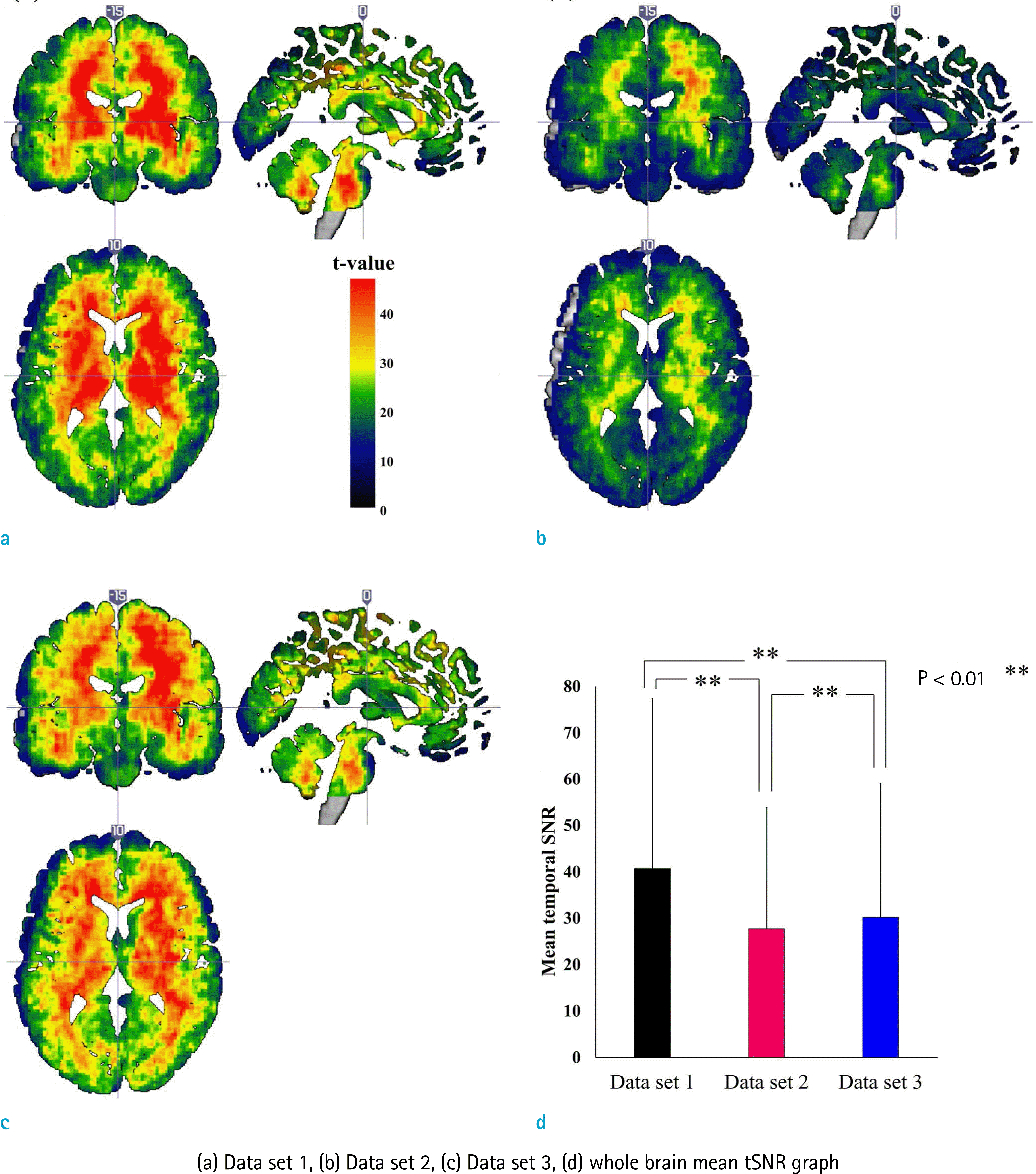
Fig. 2.
(a) One-way repeated ANOVA for the three data sets (Data set 1, Data set 2, Data set 3). The SPM{F}s had a threshold of P < 0.05, with FWE-correction for multiple comparisons at the whole brain level. Post-hoc two sample t-tests showed that the difference between Data set 1 and Data set 2 (b) and between Data set 1 and Data set 3 (c) were responsible for the difference shown in analysis of variance (ANOVA). The SPM{t}s had a threshold of P < 0.05, with FWE-correction for multiple comparisons at the whole brain level.
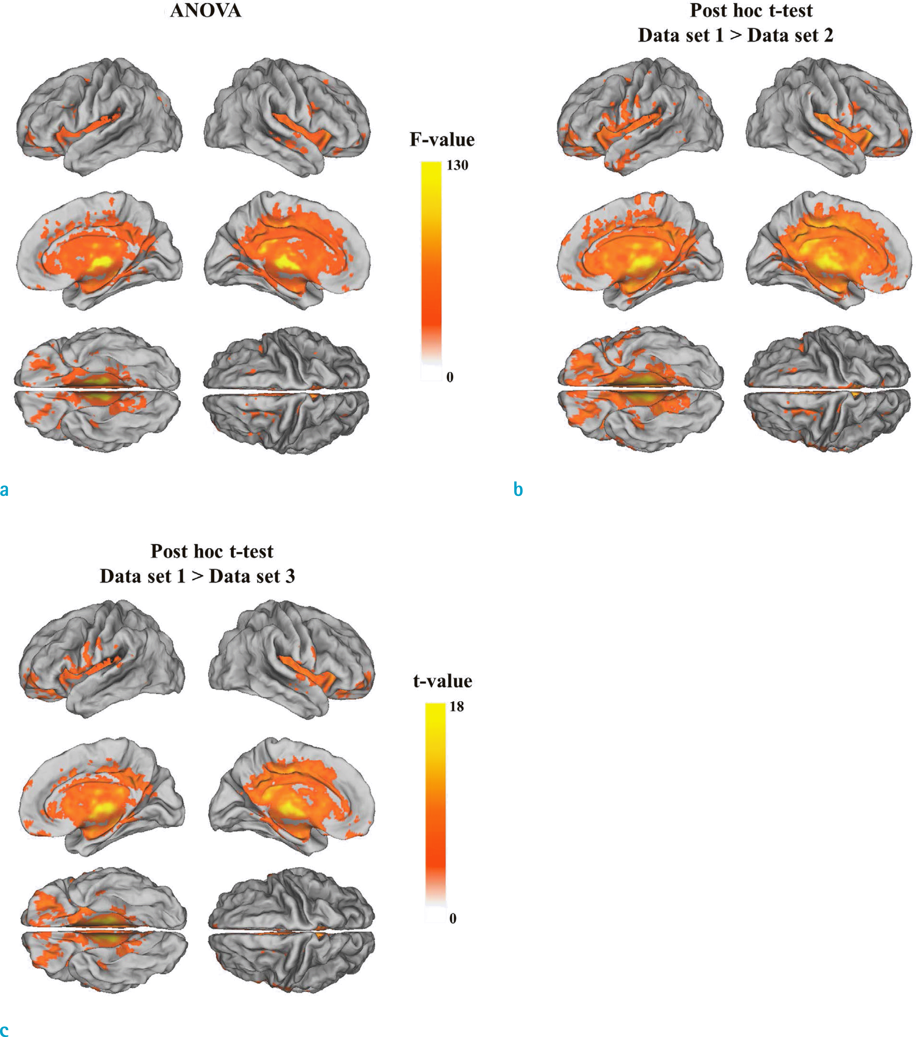
Fig. 3.
One sample t-test results of (a) default-mode networks and (b) right motor networks obtained from independent component analysis for three data sets (Data set 1, Data set 2, Data set 3). The SPM{t}s had a threshold of P < 0.05, with FWE-correction for multiple comparisons at the whole brain level. One-way repeated ANOVA for the three data sets did not show differences between data sets.

Fig. 4.
One sample t-test results of (a) default-mode networks and (b) right motor networks obtained from seed-based analysis for three data sets (Data set 1, Data set 2, Data set 3). The SPM{t}s had a threshold of P < 0.05, with FWE-correction for multiple comparisons at the whole brain level. One-way repeated ANOVA for the three data sets did not show differences between data sets.

 XML Download