

 PDF
PDF ePub
ePub Citation
 Print
Print


INTRODUCTION
Arachnoid granulations are projections of arachnoid membrane into dural sinuses, frequently located parasagitally in the transverse and superior sagittal sinuses. When arachnoid granulations are enlarged, they are termed “giant”. Previous reports have found these structures to be sometimes associated with headaches, and in some patients, idiopathic intracranial hypertension (IIH) may occur (123). The pathophysiology of these enlarged structures seen as filling defects on imaging is not clearly defined, although in some cases they are presumed to cause symptoms such as headache via pressure resulting from secondary venous sinus obstruction. We present a case of headache mimicking migraine with aura, not previously described in the setting of giant arachnoid granulations in a 39-year-old, previously healthy man.
CASE REPORT
A 39-year-old previously healthy man presented to the neurology clinic with the chief complaint of headaches described as a dull, pressure-like sensation associated with lightheadedness that began approximately three weeks prior to presentation. He reported the headaches to occur three to four days weekly on average and experienced worsening of his headaches with sleep deprivation or alcohol consumption. The headaches were not precipitated by changes in position. He reported no other associated symptoms such as visual obscurations, tinnitus or diplopia. He reported no history of previous headaches. Family history was negative for headache or other neurological disorders. Past medical history was unremarkable and he did not take any daily medications.
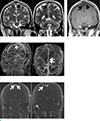
Magnetic resonance imaging (MRI) of the brain was conducted, which revealed an amorphous lesion compressing the superior sagittal sinus, shown as high signal on T2-weighted image and low signal on T1-weighted image (Fig. 1a, b). MR venography of the brain was subsequently obtained, which revealed irregular narrowing of the lumen and multifocal filling defects of the superior sagittal sinus suggestive of arachnoid granulations (Fig. 1c, d). His symptoms improved with the use of symptomatic medications including naproxen for headache and dimenhydrinate for dizziness, but he was eventually started on propranolol for headache prophylaxis. The patient stated that his headache frequency was dramatically reduced approximately three weeks following the daily use of propranolol.
DISCUSSION
Giant arachnoid granulations, thought to represent extension of endothelium-lined venous sinuses, are seen as focal ovoid masses with surrounding blood flow on neuroimaging. There structures are termed “giant” when they are sufficiently large enough to cause local dilation or filling defects. Giant arachnoid granulations are commonly seen as nonenhancing, hypointense lesions on T1-weighted MR images (4). Headaches have been reported in patients with arachnoid granulations, and imaging findings, as well as clinical presentation can lead to the differential diagnosis of venous sinus thrombosis or IIH. Although IIH is a reported manifestation, venous sinus pressure has commonly been found to be normal despite the presence of headaches, suggesting that increased pressure may not be the sole explanation for patient symptomatology (56). The headache characteristics of our patient were not indicative of venous sinus thrombosis or IIH and therefore he did not undergo further relevant evaluation. His headaches which were associated with vertigo met the International Classification of Headache Disorders (ICHD-3) criteria for migraine with aura, defined as recurrent attacks of central nervous system symptoms usually followed by headache. However, the patient did not report history of previous headaches, which is therefore suggestive of secondary headache that is migraine-like. The patient's headaches were responsive to propranolol, a widely-used medication used for prophylaxis in migraine patients. Our case is an example of headache mimicking migraine with aura in a patient with giant arachnoid granulations, a clinical manifestation not previously described in the literature. Therefore, it may be useful to consider such structural etiology in a patient presenting with headaches suggestive of migraine who reports no previous history of migraines in one's younger years.
 XML Download