

 PDF
PDF ePub
ePub Citation
 Print
Print


INTRODUCTION
Viagra is the commercial name of Sildenafil citrate, a selective phosphodiesterase type 5 (PDE-5) inhibitor. It was originally synthesized to treat hypertension and angina pectoris. However, the drug showed little effect on those clinical aspect, and was rather found to be useful for inducing penile erection. After gaining approval from Food and Drug Administration (FDA) 1998, it has been prescribed worldwide to treat male erectile dysfunction. Like many other drugs, sildenafil may cause various kinds of adverse effects. Most commonly reported adverse effects include headache (16%), facial flushing (10%), dyspepsia (7%), nasal congestion (4%), and visual disturbances (3%) (1). Sildenafil may also cause central nervous system (CNS) adverse effects such as dizziness, depression, insomnia, abnormal dreams, somnolence, and nervousness, but these are very rarely identified (12). Here we present a case of toxic encephalopathy induced by sildenafil since there were no reported cases about neuroradiologic findings of Viagra induced toxic encephalopathy.
CASE REPORT
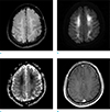
A 53-year-old man presented to our hospital with slurred speech and confused mental state. According to his older sister, he took two tablets of Viagra® (100 mg) four days earlier. He didn't have any past medical history, nor did he take any medications until then. At the time of presentation, Viagra® was the only pill he took within a month. Furthermore, the patient's sister denied the possibility of alcohol abuse or any other drug overdose. Though she couldn't remember the exact time of symptom onset, she described that his symptoms occurred after taking those tablets at noon. Clinicians evaluated his symptoms and neurologic signs thoroughly. He showed drowsy mental status and slurred speech at the time of presentation, but results of other neurologic examinations were normal. For detailed neurologic work-up, serologic tests, contrast-enhanced magnetic resonance (MR) imaging of the brain, cerebrospinal fluid (CSF) examination, and electroencephalography (EEG) evaluation were done. Serologic tests including blood sodium, glucose, osmolarity and others related with metabolic encephalopathy were within normal range. The patient's CSF examination showed no abnormalities. EEG showed generalized continuous slowing, suggestive of a severe diffuse cerebral dysfunction. No epileptiform discharges were seen. Such results generally are nonspecific, but can be most commonly observed in metabolic and toxic encephalopathy. After ruling out other possibilities clinically, the neurologist raised the possibility of toxic encephalopathy, and MRI evaluation was advised. On MRI, fluid-attenuated inversion recovery (FLAIR) image showed abnormal bilateral symmetric hyperintense signal along the periventricular white matter (Fig. 1a). The lesion showed diffusion restriction on diffusion weighted imaging (DWI) (Fig. 1b, c) without showing any enhancement on post-contrast image (Fig. 1d). Similar EEG and MRI findings can be seen in diffuse structural or degenerative processes. However, most slowly progressive neurodegenerative diseases show a normal EEG and imaging findings until late stages. In view of the patient's age and the relatively rapid onset of the neurologic symptoms, the possibility of other structural or neurodegenerative disorders were considered low. Among metabolic or toxic encephalopathy, uremic or hypoglycemic encephalopathy can show similar imaging findings. However, the patient didn't have a history of diabetes and associated lab findings were all within normal limits. So possibility of Viagra® induced toxic encephalopathy was considered the most likely diagnosis, based on the clinical situation and the imaging findings. Any other possible differential diagnoses with similar imaging and EEG findings can be excluded. Institutional Review Board approval was waived.
DISCUSSION
Viagra® is a widely used agent for the treatment of male erectile dysfunction. It is the citrate salt of sildenafil, which is a selective inhibitor of phosphodiesterase type 5 (PDE-5), an enzyme found in the corpus cavernosum (12). Its pharmacologic mechanism of action is associated with nitric oxide (NO). NO is released in the nerves and endothelial cells of penile corpus cavernosum during sexual stimulation. NO sequentially activates the enzyme called guanylate cyclase, which in turn produces cGMP. cGMP induces arterial and trabecular smooth muscle relaxation, leading to penile erection. PDE-5 is an enzyme that normally degrades cGMP. PDE-5 inhibitors such as sildenafil inhibit the degradation of cGMP by PDE-5, consequently inducing effective penile erection (34).
Like most drugs, this drug can cause various adverse events and side effects. According to the manufacturer, in pre-marketing clinical trials with over 3700 patients aged 19 to 87 years, the most common adverse events reported were headache (16%), flushing (10%), dyspepsia (7%), nasal congestion (3%), and abnormal vision (3%). Some neurologic adverse events such as ataxia, hypertonia, neuralgia, neuropathy, paresthesia, tremor, and decreased reflex had been also reported in < 2% of patients, but a causal relationship to Viagra® was uncertain. In postmarketing experience, several neurologic events like seizure, seizure recurrence, anxiety, and transient global amnesia had been reported in temporal association with Viagra® use (1). Furthermore, some published studies also have described some neurologic, psychological disturbances caused by Viagra® (2).
Although very rare, toxic encephalopathy is one of the possible neurologic adverse events of sildenafil. According to FDA, 33, 426 people reported to have side effects when taking Viagra until July 2016. Among them, 2 people (0.01%) had reported to have toxic encephalopathy (5). However, there are no reported cases about the imaging findings of Viagra-induced toxic encephalopathy until now. Also there are no available references that explain mechanism of this potentially dangerous adverse reaction of Viagra.
In our presented case, T2 and FLAIR images showed bilateral symmetrical white matter hyperintensity in periventricular area which presented diffusion restriction on DWI. Other sequences such as SWI, perfusion MR, and enhanced MR images showed no abnormalities. These similar imaging findings can be presented in other metabolic encephalopathy such as uremic or hypoglycemic encephalopathy. White matter involvement of uremic encephalopathy is extremely rare, but has been reported about 3 cases (6). Hyperglycemia in diabetic-uremic patient or direct toxicity of uremia are posed as a possible pathomechanism. In case of hypoglycemia, white matter involvement is regarded as earlier findings. Direct toxicity of aspartate are well known pathomechanism in these patients (7). Referring to other metabolic encephalopathy which showed similar imaging finding, NO related direct toxicity can be a possible pathomechanism in Viagra-induced encephalopathy. As described above, Viagra® acts by inhibiting PDE-5. Some published studies identified that Viagra® can cross blood-brain barrier (BBB). In 1989, studies proved that NO is also formed in the CNS by NO synthase (NOS), and possibly has a role in CNS transduction and function (28). It is likely that Viagra® modulates the brain function by increasing the NO-cGMP cascade. But these studies have described the relationship between aggressive behavior and hippocampal lesions, not white matter toxicity. Whether this will induce CNS adverse effect like toxic encephalopathy is unclear (2).
Our case raises some important issues. Firstly, any clinicians who frequently prescribe Viagra should have knowledge about this possible neurologic adverse effect and be careful when dealing with patients taking Viagra®, especially when they have known neurologic diseases.
 XML Download