

 PDF
PDF ePub
ePub Citation
 Print
Print


INTRODUCTION
The flexor digitorum accessorius longus (FDAL) is the most common accessory muscle in the posterior compartment of ankle area, and other accessory muscles in this area are the peroneocalcaneus internus (PCI), tibiocalcaneus internus (TCI), accessory soleus (12). The FDAL has a variable incidence of 2-14% with a male predominance, and a few bilateral cases have been reported (234567).
The FDAL may widely originate from the tibia, fibula, posterior muscle septum, leg's fascia profunda, and posterior muscular compartment and insert on the flexor digitorum longus (FDL) tendon or quadratus plantae muscle (23456). The FDAL muscle may descend between the flexor hallucis longus (FHL) tendon and neurovascular bundle or just posterolateral to the FHL tendon (246). In our case, the FDAL muscle simply descends posterolateral to the FHL tendon.
The occurrence of this accessory muscle within tarsal tunnel can cause the ankle problem, such as tarsal tunnel syndrome and FHL tenosynovitis (1237). So, the knowledge of detailed location and complication of the FDAL is important when interpreting the magnetic resonance imaging (MRI) of the ankle area.
In this report, we present a case of FDAL incidentally found on MRI.
CASE REPORT
A 35-year-old man visited the hospital for left ankle pain for following an injury after exercise. Physical examination revealed tenderness and swelling in the left ankle. X-ray showed no abnormality.
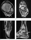
The left ankle MRI was performed using a 1.5-T MR (Achieva 1.5T SE, Philips, the Netherlands). MR findings of left ankle showed the one-headed fleshy accessory muscle arising from the posterior muscular compartment between the FHL muscle and the peroneus brevis muscle (Fig. 1). The accessory muscle descended just posterolateral to the FHL tendon, deep to the flexor retinaculum, and anterior to the Achilles tendon. In tarsal tunnel, the muscle is located lateral to neurovascular bundle and posterior to FHL tendon. Lastly, the accessory muscle inserted on the quadratus plantae muscle, and we diagnosed the accessory muscle as the FDAL. Other findings were complete tear of the posterior tibial tendon and effusion in tendon sheath.
The patient underwent posterior tibial tenorrhaphy. The accessory muscle inserting the quadratus plantae muscle was identified in surgery. After surgery, the ankle pain improved and he was followed up at regular intervals as an outpatient.
DISCUSSION
The FDAL is the second most common accessory muscle in the ankle area and the most common accessory muscle in the posterior compartment of ankle area (1). The peroneus quartus is the most common, followed by other variations such as the accessory soleus, TCI, and PCI muscles (12). The FDAL does not always bear the same morphology and it can cause clinical pathological conditions and complications (34).
This muscle was first described by Meckel, and subsequent descriptions by other authors were made, both in cadavers and live humans. According to different research groups, this muscular variation is observed in some primitive mammals, as a posterior compartment muscle of the leg (35).
The FDAL has a variable incidence of 2-14% with a male predominance, and a few bilateral cases have been reported (234567). The existence of FDAL can be associated with the abnormalities or absence of quadratus plantae muscle (5).
There are variable origin sites of the FDAL. The medial side of the tibia, the deep posterior compartment fascia of leg, and the lateral aspect of the fibula are common origin sites (2346). Among them, the most common origin of the FDAL is the fibula (3). Any structure in the posterior compartment of the leg, such as posterior muscle septum, the leg's fascia profunda, and posterior muscular compartment can be the origin sites (35).
The FDAL with one or two heads has been reported, and these one or two heads originate from different bone or fibrous points of the leg (38). Also, the FDAL muscle with one or two heads can originate from the same bone (tibia or fibula) or from both bones. The FDAL with one head is more common, and the medial head was more common than the lateral (18).
The course of FDAL muscle can be variable. In some case, the FDAL muscle may descend between the FHL tendon and neurovascular bundle, so it can be wedged shape (24). The FDAL muscle may descend just posterolateral to the FHL tendon (69). In our case, the FDAL muscle simply descends posterolateral to the FHL tendon. The course of the FDAL posterolateral to the FHL tendon is similar to the PCI, so it is difficult to differentiate the FHL from the PCI in course (6910).
The FDAL runs through the tarsal tunnel, and finally inserts on the FDL tendon or the quadratus plantae muscle. In the tarsal tunnel, the FDAL can be identified as a flesh muscle fiber at MRI, and readily distinguished from the FDL tendon (2467).
The location of the FDAL such as origin, course, and insertion can be used to differentiate the FDAL from other medial accessory muscles including the PCI, TCI, and accessory soleus muscles. The FDAL occasionally descends posterolateral to the FHL, so its course is similar to the PCI course. However, the PCI originates from only the lower fibula and inserts on the calcaneus, whereas the FDAL can originate from the tibia or posterior compartment of the leg and insert on the FDL tendon or the quadratus plantae muscle. Also, the course of the TCI is similar to the FDAL course, but the TCI inserts on the medial aspect of the calcaneus. The location of the FDAL within the tarsal tunnel and deep to the flexor retinaculum can differentiate it from the accessory soleus muscle, which runs outside of the tarsal tunnel and superficial to the flexor retinaculum (248).
Because the FDAL is related closely to the neurovascular bundle in tarsal tunnel, the occurrence of this muscle within tarsal tunnel can cause the ankle problem, such as tarsal tunnel syndrome and FHL tenosynovitis (1237). Tarsal tunnel syndrome has been reported as the most common complication in consequence of the FDAL (911).
Verification of the accessory muscles is important in foot and ankle intervention, and it result in the proper outcome. Thus, recognition and reporting of these accessory muscles are important when evaluating MRI, and detailed knowledge and evaluation of accessory muscles in the ankle region are of great importance in diagnosis and executing surgery.
 XML Download