

 PDF
PDF ePub
ePub Citation
 Print
Print


INTRODUCTION
Breast hamartomas are uncommon benign breast lesions composed of varying amounts of fatty, fibrous, and glandular elements, and are well-defined or encapsulated, separated from the rest of the glandular tissue (12). Hamartomas are not a risk factor for breast cancer development. However, there have been several reports of coexistence of carcinoma associated with hamartoma, suggesting possible malignant transformation of breast hamartomas (3). In this case report, we present a new incident of invasive ductal carcinoma (IDC) arising in a breast hamartoma.
CASE REPORT
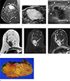
A 60-year-old woman presented with a palpable mass in her right breast. The mass had been present for several decades. One month ago, she received an abnormal result on a screening mammography which was performed at an outside hospital during a regular health check-up. She denied any history of breast surgery, trauma to either breast, or a family history of breast cancer. Physical examination revealed a 10-cm-sized soft, painless, and mobile mass in the right upper breast. Dimpling or retraction was not present. Mammography was performed at our institution and it showed a 10-cm-sized, circumscribed, irregular, fat-containing mass with a surrounding thin pseudocapsule. Within this fatty mass, in the right upper outer quadrant, a 3.5-cm-sized irregular hyperdense mass containing suspicious microcalcifications was also noted (Fig. 1a, b). Using breast ultrasonography (US), a 10-cm-sized circumscribed oval isoechoic mass was detected in the right upper breast, and an additional 3.5-cm-sized irregular hypoechoic mass was noted within the fatty mass (Fig. 1c). Suspecting breast cancer arising from a breast hamartoma, US-guided core needle biopsy using 14 gaze needle was performed, and the hypoechoic mass was proven to be an IDC. Magnetic resonance imaging (MRI) was performed preoperatively and showed a 10 cm, encapsulated fat-containing mass in the right breast suggesting breast hamartoma. And within the hamartoma, 3.5-cm-sized irregular mass with heterogeneous enhancement was noted, suggesting malignancy (Fig. 1d-f). An ipsilateral axillary lymph node was enlarged, suggesting metastasis.
Right partial mastectomy and sentinel lymph node biopsy was performed. The gross specimen revealed a large hamartoma with a smaller malignant mass which appeared as a poorly-defined, white-tan, solid mass (Fig. 1g). One ipsilateral axillary lymph node was proven to be a metastatic lymph node. The patient underwent postoperative adjuvant chemotherapy and radiation therapy and is under regular follow up observation without tumor recurrence.
DISCUSSION
Hamartoma of the breast is a rare, benign tumor, with a prevalence of < 1% of all breast lesions. The prevalence is generally underestimated because the tumor is often asymptomatic and frequently undiagnosed (4). On physical examination, hamartoma is usually a well-defined, soft, painless and mobile tumor (5). Hamartomas are often diagnosed incidentally during a screening mammography on which they show a characteristic appearance consisting of a circumscribed mass with a thin pseudocapsule and mixed fatty and soft-tissue elements (26). US features of breast hamartoma are variable, but usually appear as a well-circumscribed, oval-shaped, solid mass with internal heterogeneous echogenicity ('breast within breast appearance'). Sometimes it can be difficult to distinguish a hamartoma from the surrounding breast tissue (7). MRI shows the presence of internal fat density as well as smooth well defined hypointense rim and internal heterogeneous enhancement, which are characteristic of breast hamartoma (4).
According to several papers that reported malignancy associated with breast hamartoma, mammography mostly revealed a spiculated mass or a fatty mass containing suspicious microcalcifications within the hamartoma. On US, an irregular hypoechoic tumor within the hamartoma is the most common finding, but there are also cases with irregular hypoechoic tumors located both within and external to the hamartoma. Microscopically, hamartomas are composed of a combination of epithelial and stromal elements, usually with normal ducts or lobules (56). Malignant tumors arising from the hamartoma are known to show large tumor cells arranged in solid clusters with pleomorphic nuclei (8).
Breast hamartoma is a tumor that may be underdiagnosed by pathologists because it does not have any cytologic or architectural specificity. It contains all the constituents of normal breast tissue, and because a breast hamartoma is a benign tumor, it is not thought to be associated with breast cancer (3). Nevertheless, several reports described different types of malignancy including both non-invasive and invasive carcinomas arising in breast hamartomas. The cause of breast cancer arising from a hamartoma can be explained by the histological equivalence of its components to normal breast tissue. Case reports of carcinomas found in hamartomas suggest that malignant cells can exist in all lesions containing breast tissue. Therefore, the presence of a breast hamartoma does not exclude the possibility of breast cancer originating from this benign lesion (7).
For any lesion that shows radiologic features atypical for a hamartoma, such as suspicious additional mass with discriminating imaging features from the hamartoma or increase in size of the mass or development of microcalcifications during follow up, the possibility of breast carcinoma developing within hamartoma should be considered and further pathological diagnosis should be performed (9).
In conclusion, breast hamartomas are uncommon benign tumors and associated carcinomas occur only rarely. However, due to the possibility of coexistence of carcinoma with a breast hamartoma, we should be prudent and aware of the suspicious features within a hamartoma during the performance and interpretation of imaging studies.
 XML Download