

 PDF
PDF ePub
ePub Citation
 Print
Print


INTRODUCTION
The liver is an extranodal organ that is frequently invaded by lymphomas, with most cases involving secondary lymphomas (1). The image findings of hepatic lymphoma have been reported as variable, ranging from single or multiple nodules to diffuse infiltrates (12). It is known that hepatic lymphomas show low signal intensity on T1 weighted images, high signal intensity on T2 weighted images, and low signal intensity on hepatobiliary phase image, using hepatocyte-specific contrast agents (34). Here, we report an atypical case of hepatic lymphoma showing iso-signal intensity on hepatobiliary phase of contrast-enhanced MRI using gadoxetic acid (Gd-EOB-DTPA, Primovist; Bayer Schering Pharma, Berlin, Germany).
CASE REPORT
An 80-year-old male patient presented with fever, chills, and muscle pain for two weeks. He had a history of atrial fibrillation and had been taking warfarin. Laboratory findings were as follows: aspartate amino transferase, 338 IU/L (normal, 0-39 IU/L); alanine amino transferase, 223 IU/L (normal, 0-39 IU/L); alkaline phosphatase, 340 IU/L (normal, 30-125 IU/L); gamma-glutamyl transferase, 167 IU/L (normal, 7-66 IU/L), lactate dehydrogenase 3300 IU/L (normal, 20-480 IU/L). All values were higher than the normal range. Total bilirubin, albumin, creatinine, and prothrombin time values were within normal limit and no HIV antigen-antibody was found in an immune serum test.
Abdominal computed tomography (CT) scan showed periportal infiltrating hypodense mass and multiple round hypodense nodules in the liver. On arterial phase, there was no definite contrast enhancement. There was a gradual contrast enhancement of the peripheral portion of the lesion on portal venous and delayed phases. No abnormal bile duct dilatation or splenomegaly was observed.
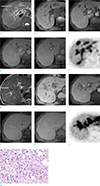
In a magnetic resonance imaging scan (Magnetom Avanto 1.5T, Siemens, Erlangen, Germany), the infiltrative lesion around the portal vein and round nodules in the liver appeared as low signal intensity on T1 weighted images (gradient-echo, TR/TE 120/2.4[out-of-phase]-4.8[in-phase] ms, flip angle 70°). The infiltrative lesion around the portal vein was slightly hypointense and the round nodules were isointense or hyperintense on T2 weighted images (respiratory-triggered turbo spin-echo, TR/TE 4807/108 ms, flip angle 140°). Meanwhile, diffusion restriction was not clear on diffusion weighted imaging. Dynamic imaging (volume interpolated gradient-echo, TR/TE 4.6/2.1, flip angle 10°, matrix of 350 × 350, 3-mm slice thickness) using Gd-EOB-DTPA revealed no definite enhancement on the arterial phase, and there was partial peripheral enhancement on portal venous phase, where the signal intensity was lower than the surrounding hepatic parenchyma. On the transitional phase, a pattern of gradual enhancement toward the center was shown, with enhancement in some parts that was similar to or slightly higher than the hepatic parenchyma. On the hepatobiliary phase, which was obtained 20 minutes after contrast injection, the border of the infiltrative lesion around the portal vein showed weak low signal intensity, and the center showed iso-signal intensity compared with the hepatic parenchyma; multiple round nodules also showed iso-signal intensity (Fig.1a-l). The probable diagnosis was IgG4 sclerotic disease with inflammatory pseudotumor, metastasis or lymphoma.
In a positron emission tomography (PET) scan, the infiltrative lesion around the portal vein and multiple round nodules in the liver showed a strong fluorodeoxyglucose (FDG) uptake. Moreover, a strong FDG uptake was shown in the adrenal gland, the fifth lumbar vertebra, the mediastinal lymph node, and the porta hepatis lymph node (Fig. 1f, l).
An ultrasound-guided biopsy was performed and the pathological diagnosis revealed necrosis in many tissue parts and lymphocytes of various sizes, having irregular nuclei. The tumor cells showed a positive reaction to CD3, TIA-1, and granzyme-B in immunohistochemistry, and the molecular pathological evaluation for the Epstein-Barr virus also showed positive results. Hence, the patient was diagnosed with nasal type extranodal NK/T-cell lymphoma (Fig.1m). The patient's condition was drastically worsened due to multiple organ failure, and he died within nine days after diagnosis, without receiving any anticancer therapy.
DISCUSSION
The liver is one of the extranodal organs frequently invaded in patients with progressive non-Hodgkin's lymphoma. Hepatic lymphoma is mostly secondary, and primary lymphoma originating in the liver is very rare. The image findings of hepatic lymphoma are variable. Primary lymphoma usually appears in one large nodule with clear boundaries, while secondary hepatic lymphoma can have a variable appearance, from one or multiple nodules to diffuse infiltration (12).
In most cases, hepatic lymphoma shows lower density than the normal hepatic parenchyma in a CT scan before contrast enhancement, and lower homogeneous contrast enhancement than the normal hepatic parenchyma after contrast enhancement (1). In MRI, most cases show low signal or iso-signal intensity on T1 weighted imaging, and moderate high signal intensity on T2 weighted imaging. Typically, lymphoma shows low signal intensity on arterial phase after contrast enhancement and homogeneous delayed enhancement in the portal venous phase, with isosignal intensity in the transitional phase (1345). Low signal intensity has been shown in the hepatobiliary phase using a hepatocyte-specific contrast agent, which has recently shown increasing access (34).
In the present case, the hepatic lymphoma showed mixed presence of an infiltrative lesion around the portal vein and multiple round nodules. CT findings were consistent with the previous studies. On MRI, all lesions showed low signal intensity on T1 weighted imaging and variable signal intensity on T2 weighted imaging. Although previous studies have reported most hepatic lymphomas to show moderate high signal intensity on T2 weighted images, Gazelle et al. (2) reported that hepatic lymphoma show isosignal intensity or heterogeneous signal intensity on T2 weighted imaging, which was consistent with our findings. The dynamic enhancement image obtained after Gd-EOB-DTPA showed delayed enhancement progressing gradually. On hepatobiliary phase, most lesions showed iso-signal intensity. All previous studies (34) reported low signal intensity on hepatobiliary phase, and none reported image findings similar to those found in this case.
Gd-EOB-DTPA is a contrast agent that has the dual characteristics of being both an extracellular fluid contrast agent as well as a hepatocyte-specific contrast agent. It is widely used in hepatic MRI. Using this contrast agent, hepatobiliary phase image can be obtained 20 minutes after injection. On hepatobiliary phase, tumors that do not have functional hepatocyte appear hypointense, as compared to surrounding enhancing liver parenchyma. Various hypothesis have been proposed regarding the mechanism of paradoxical uptake, where the signal intensity is similar to or higher than the surrounding hepatic parenchyma on hepatobiliary phase. Lee et al. (6) reported iso-signal intensity or high signal intensity of hepatocellular carcinoma may be explained by hypersecretion of bile, delayed secretion of bile due to mass effects, high preservation of hepatocyte function or maintained organic anionic transporting polypeptides (OATP). Moreover, Ha et al. (7) reported that the high signal intensity of hepatic metastases in the hepatobiliary phase can be attributed to the delayed stay of the contrast agent due to fibrosis, desmoplastic reaction, large interstitial space, and coagulative necrosis.
The nasal type extranodal NK/T cell lymphoma finally diagnosed in this case, accounts for 12% of the total non-Hodgkin's lymphoma cases that occur in Korea. The nose is the most frequently invaded organ; progress of the disease is known to be more aggressive and to have a worse prognosis than other lymphomas (8). There are few cases of invasion in other organs, barring the nose. Moreover, as far as we know, there has been no reported MRI finding of nasal type extranodal NK-T cell lymphoma with an infiltrative lesion around the portal vein and multiple nodules invading the liver, as was the case here. It is known that angiocentric invasion of the lymphoid cell is characteristic pathologic feature causing intravascular thrombosis, alongside necrosis and fibrosis (9). In this case, necrosis in the tissue was observed in the pathological finding. This may possibly have extended the time the contrast agent stayed inside the nodules, which resulted in iso-signal intensity in the hepatobiliary phase.
In addition, there was a report that the degree of the hepatic parenchymal enhancement on hepatobiliary phase in a Gd-EOB-DTPA-enhanced MRI is closely related to liver function, and that poor liver function results in weak contrast enhancement on hepatobiliary phase (10). The patient's MELD score was 7.7, the Child-Pugh score was A, and total bilirubin was within the normal range, indicating a not so severe deterioration in liver function. On hepatobiliary phase imaging that was obtained 20 minutes after contrast injection, it was observed that the contrast agent was excreted to the bile duct. However, at the time of the MRI, the amino transferase, alkaline phosphatase, gamma-glutamyl transferase, and lactate dehydrogenase of the patient were higher than the normal range. Moreover, on hepatobiliary phase imaging, the vascular structures presented as weak low signal intensity compared with hepatic parenchyma. Thus, it is possible that an inappropriate hepatobiliary phase was obtained due to decreased liver function, resulting in iso-signal intensity of the hepatic lymphoma.
In conclusion, we report a case of secondary nasal type extranodal NK/T cell lymphoma occurring in the liver, which shows iso-signal intensity on hepatobiliary phase of the contrast enhanced MRI using Gd-EOB-DTPA.
 XML Download