Abstract
Vascular tumors in the breast are rare, and most can be classified as being either angiosarcomas or hemangiomas. Hemangiomas are benign vascular tumors that are usually identified incidentally. Here, we are reporting on a case of a complex hemangioma of the breast, and describing the mammography, ultrasonography, and magnetic resonance imaging findings for this patient.
Vascular tumors in the breast are rare, and most can be classified as being either angiosarcomas or hemangiomas. Hemangiomas are benign vascular tumors that are usually identified incidentally, upon histopathological examination of lumpectomy specimens (123). To date, many cases of hemangiomas in the breast have been reported (24), and most of these case reports have focused on mammographic and ultrasonographic findings. However, there are several reports regarding magnetic resonance imaging (1456). Here, we present the case of a 47-year-old female, who had an incidentally-detected complex hemangioma of the breast, with mammography, ultrasonography, and magnetic resonance imaging findings.
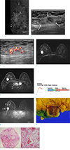
A 47-year-old woman presented with a palpable mass in the right breast that had been identified one month earlier. There was no personal medical or familial history of breast cancer. She underwent mammography and ultrasonography at an outside hospital, and was diagnosed with invasive ductal cancer in the upper outer quadrant of the right breast, after an ultrasound-guided core needle biopsy. She was referred to our hospital for surgery and underwent a preoperative re-evaluation. A right lateral spot compression magnification view (Senographe DMR; GE Healthcare, Milwaukee, WI, USA) showed a known malignant lesion as two irregular, indistinct, high density masses with microcalcifications, with a total extent of 5 cm in the upper outer portion of the right breast. Additionally, the mammography revealed a 2.5 cm, round, circumscribed, low density mass in the posterior upper direction of the known malignant lesion (Fig. 1a). There was no combined calcification within, or around, this mass.
The mass was an approximately 2.6 × 2.4 cm, oval, circumscribed, heterogeneous echoic lesion on the ultrasonography (Fig. 1b), and a power Doppler study (iU22 unit; Philips Medical System, Bothell, WA, USA) showed an increased internal vascularity (Fig. 1c). Magnetic resonance imaging (Skyra, Siemens Healthcare, Erlangen, Germany) showed a circumscribed round mass with high signal intensity on a T2-weighted image (Fig. 1d), and a dynamic contrast enhanced image showed an initial homogeneous enhancement, with delayed washout, by a gadolinium (Gd) enhanced dynamic study (Fig. 1e-f). On a maximum intensity projection (MIP) image, an irregular known malignant lesion and an incidentally detected mass with engorged vessels at the upper outer quadrant of the right breast could be observed (Fig. 1g).
The patient underwent a skin-sparing mastectomy and transverse rectus abdominus myocutaneous flap reconstruction mammoplasty. Upon gross examination, the incidentally detected tumor was found to be a spongiotic, ovoid mass with a heterogeneously dark-brown to pinkishred color and central fibrosis (Fig. 1h). Microscopically, the tumor was a circumscribed, apparently encapsulated mass with dilated, occasionally anastomosing vascular channels (Fig. 1i). These findings supported a diagnosis of complex hemangioma.
Vascular tumors of the breast are classified as either angiosarcomas or hemangiomas. Subcutaneous vascular masses are generally benign, whereas most intraparenchymal lesions prove to be malignant angiosarcomas (17). Breast hemangiomas can often be sub-classified as either cavernous, capillary, or complex hemangiomas (3). Cavernous hemangiomas are characterized by the proliferation of large dilated vessels that are separated by fibrous septae, with areas of thrombosis, infarction, and revascularization. Capillary hemangiomas are characterized by small vessels with muscular walls, and are often associated with a large feeding vessel. In complex hemangiomas, a mixture of large, thin-walled dilated vessels and small capillary-sized vessels can be observed. Grossly, hemangiomas are typically well-circumscribed brown masses that can appear to be spongy. Microscopically, the vascular channels may blend with the surrounding breast parenchyma. The dilated vessels are congested with red blood cells, separated by fibrous septa, and can exhibit extensive fibrosis, sometimes with phleboliths (3).
The mammographic findings of hemangiomas are not specific, and can appear as well-circumscribed macrolobulated lesions that may contain calcification (128). In this case, the mass was also well-circumscribed, but had no calcifications. Ultrasonographically, hemangiomas generally appear as lobulated, superficial, well-circumscribed, solid masses that are predominantly hypoechoic, and can contain areas of calcification. Additionally, they can be poorly defined, and either isoechoic or mildly hyperechoic compared with the surrounding fat, or can exhibit the features of a complex cyst (128). Our case had similar ultrasound features, including a heterogeneous echotexture, a circumscribed margin, an oval shape, and increased interval vascularity.
There are several reports of MRI findings for breast hemangiomas (12456). Hemangiomas have been reported to exhibit homogeneous hyperintensity on T2-weighted images and early intensive enhancement followed by a plateau or washout type, or slow delayed enhancement by a Gd-enhanced dynamic study (145). Kim et al. (2) reported a giant capillary hemangioma that showed slow continuous enhancement in its peripheral portion, but our complex hemangioma showed initial homogeneous enhancement with delayed washout after contrast enhancement. Differences in the vascular morphology, numbers, blood flow and tumor size might be the reason for the different curve type. Vuorela et al. (6) reported that stagnant, slowly flowing blood and thrombosis are presumably responsible for the high signal intensity on the T2-weighted images.
In summary, we reported a complex hemangioma of the breast, with the MRI finding of the initial homogeneous enhancement with delayed washout, although other MR features suggested a benign nature, such as the morphology and T2 signal intensity.
Figures and Tables
Fig. 1
47-year-old woman with a palpable mass diagnosed as invasive ductal cancer in the right breast. (a) A right lateral spot compression magnification view, showing two irregular, indistinct, high density masses with microcalcifications in the upper outer portion of the right breast, as evidenced by skin markers of biopsy-confirmed invasive ductal cancer (arrowheads). Additionally mammography revealed a 2.5 cm, round, circumscribed, low density mass (arrows) at the posterior upper direction of the known malignant lesion. There was no combined calcification within or around this mass. (b) Ultrasonography showing a 2.6 × 2.4 cm, oval, circumscribed, heterogeneous echoic mass (arrows). (c) A power Doppler study, showing increased internal vascularity in the mass. (d) A T2-weighted image showing bright, and high, signal intensity for the mass. (e, f) A dynamic contrast-enhanced image of the mass, showing initial homogeneous enhancement, with delayed washout. (g) An MIP image, showing an irregular known malignant lesion (arrowheads) and an incidentally detected mass (arrows) with engorged vessels, at the upper outer quadrant of the right breast. (h) By gross examination, the tumor (arrows) was a spongiotic, ovoid mass, with a heterogeneously dark-brown to pinkish-red color and central fibrosis. (i) Microscopically, the tumor was a circumscribed, apparently encapsulated mass (magnification × 4, left side) with dilated, occasionally anastomosing vascular channels (magnification × 200, right side).

References
1. Glazebrook KN, Morton MJ, Reynolds C. Vascular tumors of the breast: mammographic, sonographic, and MRI appearances. AJR Am J Roentgenol. 2005; 184:331–338.
2. Kim SM, Kim HH, Shin HJ, Gong G, Ahn SH. Cavernous haemangioma of the breast. Br J Radiol. 2006; 79:e177–e180.
3. Rosen PP, Hoda SA, Brogi E, Koerner FC. Rosen's breast pathology. 4th ed. Philadelphia Wolters Kluwer Health/Lippincott Williams & Wilkins;2014.
4. Yang LH, Ma S, Li QC, Xu HT, Wang X, Wang EH. A suspicious breast lesion detected by dynamic contrastenhanced MRI and pathologically confirmed as capillary hemangioma: a case report and literature review. Korean J Radiol. 2013; 14:869–873.
5. Hayasaka K, Tanaka Y, Saitoh T, Takahashi M. Gadoliniumenhanced dynamic MRI of breast hemangioma. Comput Med Imaging Graph. 2003; 27:493–495.
6. Vuorela AL. MRI of breast hemangioma. J Comput Assist Tomogr. 1998; 22:1009–1010.
7. Jozefczyk MA, Rosen PP. Vascular tumors of the breast. II. Perilobular hemangiomas and hemangiomas. Am J Surg Pathol. 1985; 9:491–503.
8. Tilve A, Mallo R, Perez A, Santiago P. Breast hemangiomas: correlation between imaging and pathologic findings. J Clin Ultrasound. 2012; 40:512–517.


 PDF
PDF ePub
ePub Citation
 Print
Print


 XML Download