

 PDF
PDF ePub
ePub Citation
 Print
Print


INTRODUCTION
Madelung's disease, or benign symmetric lipomatosis, is an uncommon disorder that is defined by the presence of multiple symmetrical fatty accumulations, which usually involve the upper trunk, neck and head (1). This disease predominantly affects men of Mediterranean origin, between the ages of 30 and 60 years, with a history of alcohol abuse. Patients usually complain of cosmetic issues, although they may also suffer from difficulties in swallowing and turning the head, and complain of neck pain or symptoms associated with the organs in the mediastinum (2). Hepatic diseases, such as cirrhosis, or fatty degenerative changes, appear in most patients (3); however, the CT and MRI findings related to Madelung's disease have not been well described. Here, we reported on an unusual case regarding a patient who presented with symmetrically located bilateral masses in the anterior neck, which had grown progressively over a period of seven years. This case was confirmed both surgically and clinically, although surgical excision was insufficient and not curative.
CASE REPORT
A 64-year-old male presented with bilaterally anterior neck masses, which had been growing progressively over a period of seven years. The patient denied any drug use, and his past medical history was unremarkable. Over a period of 30 years, the patient was a heavy alcoholic. On admission, his height was 165 cm, and his weight was 69.8 kg (BMI = 25.64, indicating obesity). At the physical examination, regions of localized swelling were observed on the neck and upper trunk, with no sign of systemic edema (Fig. 1). There was no tenderness, or palpable mass, in the chest and abdominal areas. The patient had no other neurologic abnormality. His admission laboratory findings were significant for elevated white blood cell counts (WBC) 12.6 103/uL (normal; 4.0-11.0 103/uL), aminotransferase (AST) 93 IU/L (normal; 10-40 IU/L) and alanine transferase (ALT) 108 IU/L (normal; 6-40 IU/L); other laboratory data were unremarkable. The overall appearance of the patient revealed large symmetrical, protruding mass lesions at the anterior aspects of the bilateral supraclavicular areas (Fig. 1).
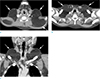
A chest radiograph (not shown) revealed hyperdense soft-tissue mass lesions at the bilateral neck and supraclavicular areas. Axial (Fig. 2a-b) and reformatted coronal (Fig. 2c) CT scans of the neck and upper chest showed diffuse hypointense lesions, located in the subcutaneous fat layer of mainly the bilaterally supraclavicular areas. These lesions were isointense, with adjacent normal fat tissue, and not separated by a normal subcutaneous fat layer. No solid portions were detected, and there were only normal vascular structures traversing within the lesion and the superficial fasciae.
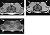
On the axial T1- (Fig. 3a), T2-weighted (Fig. 3b), fat-suppressed, and contrast-enhanced T1-weighted MRI (Fig. 3c), these lesions showed iso-signal intensity with adjacent fat at mainly the bilaterally supraclavicular areas, and did not extend into the deep fascia, the intermuscular fascia, or the intramuscular areas. They exhibited a tendency to cause outside contour bulging, rather than the displacement, compression or invasion of the adjacent normal structures. Furthermore, they had no margin or capsule, but rather appeared to be like diffuse expanding subcutaneous fat layers. As was observed in the CT scans, no solid enhancing portions that might suggest other soft-tissue tumors were seen.
Because of the cosmetic issue, surgical excision of the masses at both anterior neck areas was performed. Microscopically, the lesions were composed of normal adipose tissue, without malignant changes and atypical spindle cells, and were diagnosed as lipomas. The diagnosis of Madelung's disease was made, based on the combination of clinical, CT and MRI features.
DISCUSSION
Madelung's disease was first described in 1846, and Otto Madelung reported a series of patients with this disease in 1888 (4). This condition is characterized by massive symmetrical deposits of adipose tissue in the upper trunk, neck and head. Historically, Madelung's disease has been mainly encountered in men (male-to-female ratio = 15:1) between 30 and 60 years of age, with the disease being more prevalent among the Mediterranean population (5). Although its pathophysiology remains unclear, it is believed that Madelung's disease is related to excessive alcohol consumption, particularly that of red wine. However, despite having a history of excessive alcohol consumption, our patient had no history specifically related to red wine consumption. One study reported that, out of nine patients, only two had an association with alcohol abuse (6). On the other hand, it has been suggested that Madelung's disease might be caused by a local defect in catecholamine-induced lipolysis (7). Several other studies asserted that deletions in the mtDNA have been detected in some patients, although a defect in the mitochondrial respiratory chain could be an elusive biochemical defect (89).
Madelung's disease has two clinical phenotypes. The Type I clinical phenotype includes the male sex, and displays fat accumulation in the neck, supraclavicular, and deltoid regions. In Type II, which includes both sexes, the lipomas are distributed over much of the body, including the hips and thighs (7). Our patient was probably of the Type I clinical phenotype. Although Madelung's disease is very rare in the Asian population, the correct diagnosis of this disease is not challenging, as it can be made by reviewing the patient's clinical history, particularly with regard to alcohol consumption, and the typical imaging findings of symmetrical fat deposition.
Magnetic resonance imaging (MRI), and computed tomography (CT) can accurately demonstrate the excess fat, which has the same signal intensity and density as normal fat. An imaging study is essential before undertaking an operation, because it is important that the extent and distribution of the abnormally proliferated fat and the relations between the abnormal fat tissue and the major head and neck vessels, trachea, and esophagus be established (10). In our case, the distribution of excess fat was symmetrical and unencapsulated, and it was mainly located in the anterior subcutaneous fat layer of the supraclavicular fossa, although it is worth noting that fat deposits may be present in the neck, posterior cervical triangle, and supraclavicular fossa. MRI and CT can show the relationship between the excess fat and the surrounding great vessels, larynx, muscles and other structures, and enable the differentiation of Madelung's disease and other soft tissue tumors.
The clinical management of Madelung's disease involves abstinence from alcohol; however, there is only a slight regression in the magnitude of the lipomatous deposits (11). Surgical treatment, such as lipectomy or liposuction, is the treatment of choice. In particular, surgery is important for the symptomatic relief of patients with respiratory problems, including dyspnea and dysphagia. Because the fatty deposits of Madelung's disease are unencapsulated, and diffusely infiltrate muscles and vessels without a clear plane for dissection, complete excision is very difficult and recurrence rates are high (3).
Although Madelung's disease has characteristic imaging and clinical history findings, familial multiple lipomatosis (FML) should be carefully considered in the differential diagnosis. Clinically, FML is marked by discrete lipomas that predominate on the extremities, and are generally absent from neck and shoulders (12). Furthermore, carefully recording the family medical history can result in a diagnosis, since FML is always hereditary and usually autosomal dominant. Other diagnoses, such as: diet-related obesity, Cowden Syndrome, Proteus Syndrome, and Dercum's disease also can be ruled out, because the patient had never reported any abnormal skin lesions, dysmorphic features, or apparent developmental delays, nor did he have a history of intestinal polyps (4).
In conclusion, we present an unusual case of diffuse, bilaterally distributed fatty tissue through the supraclavicular areas, which is rare in the Asian population. Because this disease shows progressive and occasionally unpredictable behavior, it is necessary to make a correct diagnosis, not only for aesthetic reasons but also for its functional and sometimes life threatening consequences. Madelung's disease can be diagnosed without difficulty, because of its characteristic imaging and clinical findings and typical locations, although it is unusual, especially in Asia.

 XML Download