

 PDF
PDF ePub
ePub Citation
 Print
Print


INTRODUCTION
Cri-du-chat syndrome is caused by the total or partial deletion of the short arm of chromosome 5, and thus is also known as 5p-syndrome (1). The characteristic clinical features of cri-du-chat syndrome include high-pitched crying, microcephaly, facial dysmorphia, mental retardation, and developmental delay. However, high-pitched crying, one of the most characteristic features, usually disappears 1-2 years after birth. Here, we report the MRI findings of a girl with cri-du-chat syndrome and a review of the current literature.
CASE REPORT
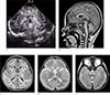
The patient was a female infant born at 382/7 weeks gestation by normal spontaneous vaginal delivery, with a birth weight of 3060 g. No significant anomalies were detected during the prenatal ultrasonography (US). She had initially been admitted to the neonatal intensive care unit the day after she was born for evaluation of oxygen desaturation during bottle-feeding. On admission, she showed high-pitched crying and abnormal facial morphology, including hypertelorism and mandibular retrognathia, which were characteristic of cri-du-chat syndrome. Cranial US on admission showed subependymal cysts at the caudothalamic groove on both sides (Fig. 1). An atrial septal defect was also detected on echocardiography at the time of admission. Ten days after admission, chromosome analysis revealed 46,XX,del (5) (p15.1), and the diagnosis of cri-du-chat syndrome was confirmed. At 3 months, the subependymal cysts were no longer visible on cranial US. On the basis of the Bayley Scales of Infant Development (second edition; BSID II), she was diagnosed with developmental delay and enrolled in a rehabilitation program at our hospital. Brain MRI at the age of 21 months revealed hypoplasia of the brain stem, a normal cerebellum, thinning of the corpus callosum, and a lack of myelination in both anterior limbs of the internal capsule (Fig. 1). Except for the anterior limbs of the internal capsule, myelination of white matter in the cerebral and cerebellar hemispheres was within normal limits.
DISCUSSION
Since cri-du-chat syndrome was first described by Lejeune et al. (2), a few radiological findings of this condition have been reported (134567). The majority of reported cases have shown hypoplasia of the brain stem, mainly involving the pons, as an associated finding of cri-du-chat syndrome (34). Other findings included atrophic middle cerebellar peduncles, atrophic cerebellar white matter, cerebellar (or vermian) hypoplasia, thinning (or dysgenesis) of the corpus callosum, reduced myelination in anterior limbs of the internal capsule, and mega cisterna magna. Similar to previous reports, in our patient, MRI at 21 months showed hypoplasia of the brain stem and thinning of the corpus callosum (3456). Thus far, reduced myelination in both anterior limbs of the internal capsule has only been reported in a 16-month-old girl who presented with brain stem hypoplasia, mild thinning of the corpus callosum, and mega cisternal magna (5). We suggest that the lack of myelination in the anterior limbs of the internal capsule could be considered one of the radiological findings associated with cri-du-chat syndrome. However, we currently do not have an explanation for this finding, and were unable to find any literature reporting an association between myelination of white matter and cri-du-chat syndrome. We hypothesize that decreased myelination in the anterior limbs of the internal capsule plays a role in the developmental delay associated with cri-du-chat syndrome. However, further study is needed to explain the clinical significance of this finding.
In addition, our patient exhibited bilateral subependymal cysts 2 days after birth. To our knowledge, an association of neonatal subependymal cysts with cri-du-chat syndrome has not been reported. There have been a few reported cases of cri-du-chat syndrome diagnosed antenatally, owing to the presence of choroid plexus cysts and other abnormal findings, such as cerebellar hypoplasia, nasal bone hypoplasia, or nuchal skin edema (8).
In conclusion, we report the radiological findings of a patient with cri-du-chat syndrome. We believe that these radiological findings, specifically brain MRI, could be a diagnostic clue for cri-du-chat syndrome in children with or without characteristic clinical features of this condition.
 XML Download