

 PDF
PDF ePub
ePub Citation
 Print
Print


Unilateral renal cystic disease (URCD) is a multicystic disease, characterized by the replacement of one kidney by multiple cysts of varying sizes without the association of cysts in the contralateral kidney.1 The disease is non-familial, non-progressive, and not related to autosomal dominant polycystic kidney disease (ADPKD).1 Except for its unilateral localization, the gross and histological findings of URCD are indistinguishable from those of ADPKD. However, unlike ADPKD, there are no genetic background and renal function deterioration in URCD patients.2
URCD is rare, so far 54 cases have been reported for 42 years since 1964.3 However, there are only 25 cases which are pathologically diagnosed and the other cases are diagnosed by imaging only.4 This study reports a case of pathologically diagnosed URCD, together with literature reviews.
CASE REPORT
A 31-year-old woman presented with chronic, intermittent, lancinating pain in the left flank area for 1 year duration. There were no gross hematuria, fever, and lower urinary tract symptoms. And other cardiovascular, respiratory, gastrointestinal, and neurologic symptoms were also absent. No family illnesses including renal diseases were reported. Upon physical examination, blood pressure was 120/80 mmHg. Left flank area was normally palpated. No other abnormalities were found.
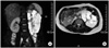
Urinalysis was normal with specific gravity of 1.012. Serum creatinine level was 0.7 mg/dl and other blood tests showed normal findings. 24 hours urine creatinine clearance was 70.11 ml/min. Abdominal enhanced computed tomography (CT) revealed enlarged left kidney filled with variable-sized multiple cysts with enhancing normal renal tissue between the cysts. However, there were no cysts in the right kidney and other intra-abdominal organs such as liver, pancreas, or spleen. Because the patient had chronic irritating pain, and we could not exclude the possibility of malignancy, radical nephrectomy was planned. For identifying clear anatomical structures for surgery, magnetic resonance imaging (MRI) was checked. A gadolinium-enhanced MRI revealed enlarged (14.0×10.0×8.0 cm) left kidney entirely filled with multiple round, well-marginated cysts of varying size without capsule formation (Fig. 1). There were no solid portions within the cysts and normally enhancing renal parenchyma between the cysts.
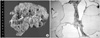
Laparoscopic radical nephrectomy was performed via transperitoneal approach. Left kidney was huge and easily dissected without adhesions. Grossly the surgical specimen measured 13.0×8.0×6.0 cm and weighed 320 g. Renal parenchyma was almost replaced by varying-sized cysts containing clear serous fluid and focally remaining parenchymal tissues were seen as thick reddish septae of cysts (Fig. 2A). Histological examination of the specimen showed multiple cysts of variable size and focal thickening of septae containing glomeruli and tubules. The cyst wall was focally lined by a layer of flattened epithelial cells. Some cyst walls showed an attached glomerulus revealing involvement of Bowman's space (Fig. 2B).
Based on the clinical features, imaging and pathological findings, and absence of a family history of renal disease, the patient was diagnosed as URCD. The patient is on outpatient-based follow-up and maintains a good renal function.
DISCUSSION
Unilateral renal cystic disease (URCD) is a rare and poorly understood condition, first described in 1964 as 'unilateral polycystic renal disease'.3 Other terms used for the same condition are localized or segmental cystic disease of the kidney (LSCDK), multiple unilateral renal cysts, segmental polycystic kidney disease, and unilateral polycystic renal disease.5 Based on the table of classification of renal cystic diseases, URCD or LSCDK is classified as a distinct entity of renal cystic diseases and the terms 'unilateral renal cystic disease (URCD)' and 'localized or segmental cystic disease of the kidney (LSCDK)' are most commonly used terms at present.5 On a review of the world literature, 54 cases of URCD were reported so far and only 25 cases of them were diagnosed histologically.4
The morphologic feature of URCD is indistinguishable from that of ADPKD both radiologically and pathologically. However URCD has five aspects different from ADPKD: (1) unilateral localization, (2) negative family history, (3) no progression to chronic renal failure, (4) no cysts in other intra-abdominal organs such as liver, spleen, or pancreas, and (5) no disorders affecting other body organ systems.2
The pathogenesis of URCD is obscure, although an acquired mal-developmental origin is hypothesized.4 Simple cysts can appear coincidentally in the contralateral kidney.5 And URCD is not distinguishable in its morphology from multiple simple cysts radiologically or pathologically only. Thus, the proposal that URCD may represent a variation in the theme of multiple simple cysts has been offered.6
Most patients are usually diagnosed in adulthood, although young people or children and even infants and neonates are on record.4 Slywotzky and Bosniak7 reported that there appears to be a male predilection. The most common clinical symptoms are flank or abdominal pain, a palpable mass, gross or microscopic hematuria, and hypertension with well-preserved renal function.4
Because URCD is characterized by replacement of various amounts of one kidney by multiple cysts, the radiographic findings depend on the number and size of the cysts and the amount of normal renal parenchyma between the cysts.7 CT and MRI best reveal the salient imaging characteristics of URCD. Variably sized cysts are separated by normally enhancing renal parenchyma. The parenchyma may become quite attenuated depending on the size and proximity of the neighboring cysts and can be misinterpreted as enhancing septae.7 Kohno and Yunoki1 reported that two patterns of involvement are noted on imaging studies. In the diffuse form, multiple cysts are scattered throughout the affected kidney, and in the segmental form, the cysts predominate in one region of the involved kidney. However, in either form, no distinct encapsulated renal mass is formed unlike multilocular cystic nephroma or cystic carcinoma.7 Pathologically URCD involves part to most or (more rarely) the entirety of one kidney with many to innumerable, variously sized cysts separated in places by normal or compressed renal parenchyma, an overall picture that recalls that of ADPKD.4
 XML Download