

 PDF
PDF ePub
ePub Citation
 Print
Print


Wilms' tumor is an extremely rare malignant renal tumor in adults, and usually presents as a parenchymal mass resembling renal cell carcinoma (RCC). In the literature, fewer than ten cases manifesting as a renal pelvis tumor have been reported, and all of these cases developed in only pediatric patients.1 Thus, our case is first reported case of intrapelvic Wilms' tumor mimicking a renal pelvis tumor in an adult.
CASE REPORT
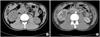
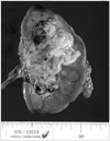
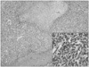
A 24-year-old man presented with painless gross hematuria. A computed tomography (CT) scan revealed a 6cm-sized solid mass located at the pelvis of the right kidney with no evidence of lymph nodal enlargement or abdominal organ metastasis (Fig. 1). Additional chest X-ray and bone scan showed no abnormal lesion. Although cystoscopic examination and urine cytology produced no abnormal findings, CT findings including tumor location and contrast enhancement pattern were highly suggestive of malignant pelvis tumor rather than benign tumor. The preoperative diagnosis was renal pelvis tumor, possibly, cT2-3N0M0, transitional cell carcinoma and radical nephroureterectomy with bladder cuff excision was performed. Grossly, a well-demarcated mass (6.0×6.0×3.5cm) was found in the renal pelvis with a grayish white, and lobulated, cut surface (Fig. 2). In contrast to the preoperative diagnosis, the histopathologic examination revealed a Wilms' tumor comprised of undifferentiated blastemal cells (90%) and focal stromal components (10%) (Fig. 3). The tumor extended to the renal cortex about 0.5cm distant from the renal capsule and final diagnosis was biphasic type Wilms' tumor, stage I. The patient received 6 cycles of vincristine plus actinomycin chemotherapy based on the National Wilms' Tumor Study-5 (NWTS-5). The patient remains healthy and was without evidence of tumor recurrence on a follow-up CT scan at 18 months postoperatively.
DISCUSSION
Wilms' tumor is the most common pediatric renal tumor but it is rare in adults; fewer than 300 adult Wilms' tumor cases have been reported in the literature.2-4 Moreover, the preoperative diagnosis of Wilms' tumor in adults is difficult, because its clinical manifestations and radiologic findings are indistinguishable from those of renal cell carcinoma, which is the most common adult renal neoplasm.5
Interestingly, in the present case, clinical and radiologic features suggested a renal pelvis tumor rather than RCC. In 1976, Engel described a Wilms' tumor in a child that completely filled the kidney collecting system, which led to loss-of-function of the involved kidney.6 To our knowledge, fewer than 10 cases manifesting as a renal pelvic mass filling the primary collecting system have been reported and only one such case has been previously reported in Korea in a 6-year-old pediatric patient.1,7 In contrast with previous pediatric cases, our patient represents the first reported case of an intrapelvic Wilms' tumor in an adult patient. The clinical presentation of intrapelvic Wilms' tumor differs in some respects from the classical form. The most common presenting symptom in cases of intrapelvic Wilms' tumor is hematuria; 87.5% of patients have hematuria at initial presentation compared with only 25% of patients with the classical form.8 Therefore, intrapelvic Wilms' tumors appear to resemble renal pelvis tumors rather than RCC in terms of their clinical features as well radiologic findings. Radical nephroureterectomy is the recommended surgical treatment for an intrapelvic Wilms' tumor, especially when the preoperative diagnosis is uncertain.9 Reziciner et al. 10 reported that one patient with an intrapelvic Wilms' tumor experienced recurrence in the ureteral stump 3 months after radical nephrectomy alone, and recommended that bladder cuff excision should be performed because transitional cell carcinoma could not be ruled out.
The experience gained during the diagnosis and treatment of intrapelvic Wilms' tumor in the present case alerted us to the need to include Wilms' tumor in the differential diagnosis of adults presenting with a renal pelvis tumor.
 XML Download