

 PDF
PDF ePub
ePub Citation
 Print
Print


Abstract
Purpose
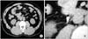
Non-enhanced spiral computed tomography (NESCT) has become the preferred method for imaging urinary calculi. We evaluated the Hounsfield units (HU) of urinary calculi on NESCT as a predictor of the therapeutic effect of extracorporeal shockwave lithotripsy (ESWL).
Materials and Methods
We evaluated 72 patients with urinary calculi who underwent ESWL. The HUs of calculi were measured on the pretreatment NESCT and at post-treatment via radiographic assessment. The patients were divided into 2 groups, the response group (n=39): the patients with remnant calculi less than 3mm or they were without remnant calculi, and the non-response group (n=33): the patients with remnant calculi greater than 3mm. The HUs of the response and non-response groups were then compared.
Figures and Tables
References
1. Sierakowski R, Finlayson B, Landes RR, Finlayson CD, Sierakowski N. The frequency of urolithiasis in hospital discharge diagnoses in the United States. Invest Urol. 1978. 15:438–441.
2. Leusmann DB, Niggemann H, Roth S, von Ahlen H. Recurrence rates and severity of urinary calculi. Scand J Urol Nephrol. 1995. 29:279–283.
3. Ahn SS, Lee SH, Kang IM. The value of non-enhanced spiral CT in the diagnosis of suspected urolithiasis. Korean J Urol. 2002. 43:1008–1013.
4. Smith RC, Verga M, McCarthy S, Rosenfield AT. Diagnosis of acute flank pain: value of unenhanced helical CT. AJR Am J Roentgenol. 1996. 166:97–101.
5. Smith RC, Verga M, Dalrymple N, McCarthy S, Rosenfield AT. Acute ureteral obstruction: value of secondary signs of helical unenhanced CT. AJR Am J Roentgenol. 1996. 167:1109–1113.
6. Levine JA, Neitlich J, Verga M, Dalrymple N, Smith RC. Ureteral calculi in patients with flank pain: correlation of plain radiography with unenhanced helical CT. Radiology. 1997. 204:27–31.
7. Olcott EW, Sommer FG, Napel S. Accuracy of detection and measurement of renal calculi: in vitro comparison of three-dimensional spiral CT, radiography, and nephrotomography. Radiology. 1997. 204:19–25.
8. Narepalem N, Sundaram CP, Boridy IC, Yan Y, Heiken JP, Clayman RV. Comparison of helical computerized tomography and plain radiography for estimating urinary stone size. J Urol. 2002. 167:1235–1238.
9. Mostafavi MR, Ernst RD, Saltzman B. Accurate determination of chemical composition of urinary calculi by spiral computerized tomography. J Urol. 1998. 159:673–675.
10. Nakada SY, Hoff DG, Attai S, Heisey D, Blankenbaker D, Pozniak M. Determination of stone composition by noncontrast spiral computed tomography in the clinical setting. Urology. 2000. 55:816–819.
11. Saw KC, McAteer JA, Fineberg NS, Monga AG, Chua GT, Lingeman JE, et al. Calcium stone fragility is predicted by helical CT attenuation values. J Endourol. 2000. 14:471–474.
12. Chaussy C, Schuller J, Schmiedt E, Brandl H, Jocham D, Liedl B. Extracorporeal shock-wave lithotripsy (ESWL) for treatment of urolithiasis. Urology. 1984. 23:59–66.
13. Singal RK, Denstedt JD. Contemporary management of ureteral stones. Urol Clin North Am. 1997. 24:59–70.
14. Politis G, Griffith DP. ESWL. Stone-free efficacy based upon stone size and location. World J Urol. 1987. 5:225–228.
15. Mattelaer P, Schroder T, Fischer N, Jakse G. In situ extracorporeal shockwave lithotripsy of distal ureteral stones: parameters for therapeutic success. Urol Int. 1994. 53:87–91.
16. Bon D, Dore B, Irani J, Marroncle M, Aubert J. Radiographic prognostic criteria for extracorporeal shock-wave lithotripsy: a study of 485 patients. Urology. 1996. 48:556–560.
17. Federle MP, McAninch JW, Kaiser JA, Goodman PC, Roberts J, Mall JC. Computed tomography of urinary calculi. AJR Am J Roentgenol. 1981. 136:255–258.
18. Joseph P, Mandal AK, Singh SK, Mandal P, Sankhwar SN, Sharma SK. Computerized tomography attenuation value of renal calculus: Can it predict successful fragmentation of the calculus by extracorporeal shock wave lithotripsy? A preliminary study. J Urol. 2002. 167:1968–1971.
19. Gupta NP, Ansari MS, Kesarvani P, Kapoor A, Mukhopadhyay S. Role of computed tomography with no contrast medium enhancement in predicting the outcome of extracorporeal shock wave lithotripsy for urinary calculi. BJU Int. 2005. 95:1285–1288.
20. Pareek G, Armenakas NA, Panagopoulos G, Bruno JJ, Fracchia JA. Extracorporeal shock wave lithotripsy success based on body mass index and Hounsfield units. Urology. 2005. 65:33–36.
21. Byeon SS, Jeon SS, Lee HW, Park EC, Lee JH, Kwak C, et al. Ureteroscopic manipulation for ureteral calculi: comparison with ESWL. Korean J Urol. 1996. 37:1124–1131.
22. Erturk E, Herrman E, Cockett AT. Extracorporeal shock wave lithotripsy for distal ureteral stones. J Urol. 1993. 149:1425–1426.
23. Kim SS, Sung BM, Ahn SH. Comparison of shock wave lithotripsy (SWL) and rigid ureteroscopic stone removal (URS) for treatment of upper ureteral stones. Korean J Urol. 2004. 45:444–448.
24. Dretler SP, Spencer BA. CT and stone fragility. J Endourol. 2001. 15:31–36.
 XML Download