

 PDF
PDF ePub
ePub Citation
 Print
Print


Abstract
Materials and Methods
We retrospectively reviewed the medical records and imaging studies of 33 children (21 boys, 12 girls) treated for an urachal anomaly over a ten-year period.
Results
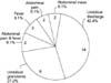
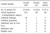
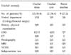
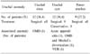
Twenty-four patients were equally diagnosed with either an urachal sinus or urachal cyst; the other nine patients were confirmed to have a patent urachus. Umbilical discharge (14 patients) and umbilical granuloma (9 patients) were the most common presentations. The 12 patients with an urachal sinus underwent ultrasonography (USG) (10; diagnostic), 2 fistulography (all; diagnostic). Those with an urachal cyst underwent either USG (6/12; diagnostic), computed tomography (CT) (3; diagnostic), fistulography (2; diagnostic), or magnetic resonance imaging (MRI) (1; diagnostic). One subject was affected by acute appendicitis, which was confirmed by CT. Of the 9 children with patent urachus, 7 underwent USG (all; diagnostic); exploration without further imaging studies was performed on the remaining 2 subjects. Surgical excision was performed in 30 patients. Omphalomesenteric duct or Meckel's diverticulum were incidental findings, which were simultaneously repaired. Conservative treatment was successful in only 3 patients.
Conclusions
Urachal anomalies in children mainly manifest as umbilical discharges and umbilical granuloma, but may present non-specific symptoms in some cases. USG is a useful method for diagnosis, but other imaging modalities can be useful to establish the differential diagnosis. A limited number of children with urachal anomalies, mainly presenting with an umbilical discharge, can be managed conservatively. However, complete surgical excision of the lesion, with the possible associated anomalies, should be the basic scheme for children with urachal anomalies.
References
1. Gearhart JP. Walsh PC, Retik AB, Vaughan ED, Wein AJ, Kavoussi LR, Novick AC, editors. Exstrophy, epispadias, and other bladder anomalies. Campbell's urology. 2002. 8th ed. Philadelphia: Saunders;2136–2196.
2. McCollum MO, Macneily AE, Blair GK. Surgical implications of urachal remnants: presentation and management. J Pediatr Surg. 2003. 38:798–803.
3. Mesrobian HG, Zacharias A, Balcom AH, Cohen RD. Ten years of experience with isolated urachal anomalies in children. J Urol. 1997. 158:1316–1318.
4. Heo JM, Yoon JB. Congenital patent urachus: report of 2 cases. Korean J Urol. 1994. 35:1151–1155.
5. Kang MS, Lee SW, Oh JS, Shin MK, Shin DM. A case of patent urachus. Korean J Urol. 1989. 30:262–265.
6. So MW, Kim HJ, Kim BK. A case of vesicourachal diverticulum. Korean J Urol. 1989. 30:266–269.
7. Yim CS, Kim ME, Lee JH, Chang DS. A case of congenital patent urachus. Korean J Urol. 1982. 23:715–717.
8. Choi BN, Lim CK, Na HJ, Song CK, Ryu SB, Kim EH. A case of congenital patent urachus. Korean J Urol. 1980. 21:373–375.
9. Bauer SB, Retik AB. Urachal anomalies and related umbilical disorders. Urol Clin North Am. 1978. 5:195–211.
10. Cilento BG Jr, Bauer SB, Retik AB, Peters CA, Atala A. Urachal anomalies: defining the best diagnostic modality. Urology. 1998. 52:120–122.
11. Leyson JF. Calcified urachal cyst. Br J Urol. 1984. 56:438.
12. Diehl K. A rare case of urachal calculus. Br J Urol. 1991. 67:327–328.
13. Davidson BR, Brown NJ, Neoptolemos JP. Haemorrhage into a urachal cyst presenting as an 'acute abdomen'. Postgrad Med J. 1987. 63:493–494.
14. Agatstein EH, Stabile BE. Peritonitis due to intraperitoneal perforation of infected urachal cysts. Arch Surg. 1984. 119:1269–1273.
15. Savanelli A, Cigliano B, Esposito G. Infected and ruptured urachal cyst causing peritonitis. Z Kinderchir. 1984. 39:267–268.
16. Nair KP. Mucous metaplasia and rupture of urachal cyst as a rare cause of acute abdomen. Br J Urol. 1987. 59:281–282.
17. Valda V, Conn MJ. Spontaneous rupture of a noninfected urachal cyst. J Pediatr Surg. 1991. 26:747–748.
18. Rich RH, Hardy BE, Filler RM. Surgery for anomalies of the urachus. J Pediatr Surg. 1983. 18:370–372.
19. Lane V. Congenital patent urachus associated with complete (hypospadiac) duplication of the urethra and solitary crossed renal ectopia. J Urol. 1982. 127:990–991.
20. Huang CS, Luo CC, Chao HC, Chen HM, Chu SM. Urachal anomalies in children: experience at one institution. Chang Gung Med J. 2003. 26:412–416.
21. Beck AD, Gaudin HJ, Bonham DG. Carcinoma of the urachus. Br J Urol. 1970. 42:555–562.
 XML Download