

 PDF
PDF ePub
ePub Citation
 Print
Print


Abstract
Purpose
We retrospectively reviewed cases of ureteroscopic lithotripsy where a pneumatic lithotriptor had been used, and report on the success and complications related to this procedure.
Materials and Methods
Between October 1996 and September 2005, ureteroscopic lithotripsy was performed in 319 cases. The medical records of 274 of these cases were available for review. The ureteroscopic lithotripsy had been performed using a rigid ureteroscope (Stortz, 10Fr) and pneumatic lithotriptor (Swiss lithoclast). A successful procedure was defined as the absence of a residual stone larger than 2mm in size on postoperative KUB or ultrasonography.
Results
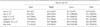
53, 32 and 189 stones were located in the upper, middle and lower ureter, which were defined as groups 1, 2 and 3, respectively. The overall success rate was 92.7%. The success rates of upper, middle and lower ureteral stones were 77.4 (41/53), 93.8 (30/32) and 96.8% (183/189), respectively. The success rate in group 1 was significantly lower than the other two groups (p<0.05). The most common cause of failure was the upward migration of the stone. The rates of stent indwelling were 37.7 (20/53), 34.5 (11/32) and 32.8% (62/189) in groups 1, 2 and 3, respectively. Perforation rates were 5.7 (3/53), 3.1 (1/32) and 2.1% (4/189) in groups 1, 2 and 3, respectively. All patients with a ureteral perforation were successfully treated with a double-J stent indwelling only for a period of 4-6 weeks. The most common complications were pain and gross hematuria.
Figures and Tables
References
1. Chaussy CG, Fuchs GJ. Current state and future developments of noninvasive treatment of human urinary stones with extracorporeal shock wave lithotripsy. J Urol. 1989. 141:782–789.
2. Lyon ES, Banno JJ, Schoenberg HW. Transurethral ureteroscopy in men using juvenile cystoscopy equipment. J Urol. 1979. 122:152–153.
3. Vorreuther R, Engelking R. Features and acoustic output of five electrohydaulic lithotriptors for endoureteral stone treatment. J Endourol. 1992. 6:41–45.
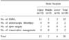
4. Fuchs GJ, Chaussy CG, Stenzl A. Current management concepts in the treatment of ureteral stones. J Endourol. 1988. 2:117–121.
5. Watson G, Murray S, Dretler SP, Parrish JA. The pulsed dye laser for fragmenting urinary calculi. J Urol. 1987. 138:195–198.
6. Denstedt JD, Eberwein PM, Singh RR. The swiss lithoclast: a new device for intracorporeal lithotripsy. J Urol. 1992. 148:1088–1090.
7. Jeon SH, Lee SJ, Lee CH, Chang SG, Kim JI. The comparison of treatment results of ureteroscopic lithotripsy with lithoclast and electrohydraulic lithotripsy. Korean J Urol. 1999. 40:542–545.
8. Sun YB, Heo DS, Woo JH, Kim YH, Park HJ, Kwon CH. 376 cases of ureteroscopic stone removal. Korean J Urol. 1999. 40:546–550.
9. Sozen S, Kupeli B, Tunc L, Senocak C, Alkibay T, Karaoglan U, et al. Management of ureteral stones with pneumatic lithotripsy: report of 500 patients. J Endourol. 2003. 17:721–724.
10. Aghamir SK, Mohseni MG, Ardestani A. Treatment of ureteral calculi with ballistic lithotripsy. J Endourol. 2003. 17:887–890.
11. Aridogan IA, Zeren S, Bayazit Y, Soyupak B, Doran S. Complications of pneumatic ureterolithotripsy in the early postoperative period. J Endourol. 2005. 19:50–53.
12. Robert M, Bennani A, Guiter J, Averous M, Grasset D. Treatment of 150 ureteric calculi with the lithoclast. Eur Urol. 1994. 26:212–215.
13. Lee KW, Lee TY, Woo YN. The experience of ureterorenoscopic lithotripsy with insertion of ureterorenoscope between two safety guide wires serving as an access port. Korean J Urol. 2003. 44:221–226.
14. Desai MR, Patel SB, Desai MM, Kukreja R, Sabnis RB, Desai RM, et al. The Dretler stone cone: a device to prevent ureteral stone migration-the initial clinical experience. J Urol. 2002. 167:1985–1988.
15. Hosking DH, McColm SE, Smith WE. Is stenting following ureteroscopy for removal of distal ureteral calculi necessary? J Urol. 1999. 161:48–50.
16. Rane A, Cahill D, Larner T, Saleemi A, Tiptaft R. To stent of not to stent? That is still the question. J Endourol. 2000. 14:479–481.
17. Denstedt JD, Wollin TA, Sofer M, Nott L, Weir M, D'A Honey RJ. A prospective randomized controlled trial comparing nonstented versus stented ureteroscopic lithotripsy. J Urol. 2001. 165:1419–1422.
18. Santa-Cruz RW, Leveillee RJ, Krongrad A. Ex vivo comparison of four lithotripters commonly used in the ureter: What does it take to perforate? J Endourol. 1998. 12:417–422.
19. Puppo P, Ricciotti G, Bozzo W, Introini C. Primary endoscopic treatment of ureteric calculi. A review of 378 cases. Eur Urol. 1999. 36:48–52.
20. Ceylan K, Sunbul O, Sahin A, Gunes M. Ureteroscopic treatment of ureteral lithiasis with pneumatic lithotripsy: analysis of 287 procedures in a public hospital. Urol Res. 2005. 33:422–425.
21. Weizer AZ, Auge BK, Silverstein AD, Delvecchio FC, Brizuela RM, Dahm P, et al. Routine postoperative imaging is important after ureteroscopic stone manipulation. J Urol. 2002. 168:46–50.
 XML Download