

 PDF
PDF ePub
ePub Citation
 Print
Print


Abstract
Purpose:
To investigate the prevalences of anatomical variations regarding prostate and its surrounding structures, and also the intra- and postoperative effects of such anatomical variations in performing radical retropubic prostatectomy (RRP).
Materials and Methods:
A retrospective analysis of 156 patients who received RRP for prostate cancer was performed. Patients’ records including the results preoperative radiologic evaluations were reviewed. For our analysis, patients were grouped according to the anatomical variations relevant to prostate and surrounding structures shown on preoperative radiographs. Also, patients were interviewed via telephone as needed.
Results:
Prostate volume (mean: 41.4ml) measured from preoperative transrectal ultrasound correlated with estimated blood loss (EBL) during RRP (p=0.029). Interspinous diameter (mean: 1.69cm) measured on axial image of preoperative magnetic resonance imaging (MRI) was observed to be inversely correlated with operative time (p=0.010). And, patients with box-shaped deep dorsal vein (as demonstrated on axial view of MRI; 15.3%) were observed to have significantly less EBL during RRP (p=0.030). Also, EBL was significantly higher (p=0.013) for patients in which anterior portion of prostatic apex appeared to overlap and obscure membranous urethra (62.8%). Meanwhile, absence of the distal protrusion of apical region (21.8%) was observed to be associated with early (within 3 postoperative months) recovery of urinary continence (p=0.014).
Conclusions:
Our results suggest that various anatomical variations regarding prostate and its surrounding structure may exist as herein presented, and also that they may indeed have significant effects on both intra- and postoperative course regarding RRPs. Variations in the shape of prostatic apex may be significantly associated with recovery of continence after RRPs. (Korean J Urol 2006;47:568-577)
Go to : 
REFERENCES
1.Headquarter of Korea Central Cancer Registry. Cancer registry system in Korea. http://www.ncc.re.kr.
2.Myers RP., Cahill DR., Devine RM., King BF. Anatomy of radical prostatectomy as defined by magnetic resonance imaging. J Urol. 1998. 159:2148–58.

3.Myers RP. Practical surgical anatomy for radical prostatectomy. Urol Clin North Am. 2001. 28:473–90.
4.Coakley FV., Eberhardt S., Wei DC., Wasserman ES., Heinze SB., Scardino PT, et al. Blood loss during radical retropubic prostatectomy: relationship to morphologic features on preoperative endorectal magnetic resonance imaging. Urology. 2002. 59:884–8.
5.Hammerer P., Hubner D., Gonnermann D., Huland H. Perioperative and postoperative complications of pelvic lymphadenectomy and radical prostatectomy in 320 consecutive patients. Urologe A. 1995. 34:334–42.
6.Rainwater LM., Segura JW. Technical consideration in radical retropubic prostatectomy: blood loss after ligation of dorsal venous complex. J Urol. 1990. 143:1163–5.
7.Hsu El., Hong EK., Lepor H. Influence of body weight and prostate volume on intraoperative, perioperative, and postoperative outcomes after radical retropubic prostatectomy. Urology. 2003. 61:601–6.
8.Goh M., Kleer CG., Kielczewski P., Wojno KJ., Kim K., Oesterling JE. Autologous blood donation prior to anatomical radical retropubic prostatectomy: Is it necessary? Urology. 1997. 49:569–73.
9.Fowler FJ Jr., Barry MJ., Lu-Yao G., Roman A., Wasson J., Wennberg JE. Patient-reported complications and follow-up treatment after radical prostatectomy. The National Medicare Experience: 1988-1990 (updated June 1993). Urology. 1993. 42:622–9.
10.Coakley FV., Eberhardt S., Kattan MW., Wei DC., Scardino PT., Hricak H. Urinary continence after radical retropubic prostatectomy: relationship with membranous urethral length on preoperative endorectal magnetic resonance imaging. J Urol. 2002. 168:1032–5.
11.Wei JT., Dunn RL., Marcovich R., Montie JE., Sanda MG. Prospective assessment of patient reported urinary continence after radical prostatectomy. J Urol. 2000. 164:744–8.
12.Steiner MS. Anatomic basis for the continence-preserving radical retropubic prostatectomy. Semin Urol Oncol. 2000. 18:9–18.
13.Strasser H., Frauscher F., Helweg G., Colleselli K., Reissigl A., Bartsch G. Transurethral ultrasound: evaluation of anatomy and function of die rhabdosphincter of the male urethra. J Urol. 1998. 159:100–4.
14.Lepor H., Kaci L. The impact of open radical retropubic prostatectomy on continence and lower urinary tract symptoms: a prospective assessment using validated self-administered outcome instruments. J Urol. 2004. 171:1216–9.
15.Karam I., Moudouni S., Droupy S., Abd-Alsamad I., Uhl JF., Delmas V. The structure and innervation of the male urethra: histological and immunohistochemical studies with three-dimensional reconstruction. J Anat. 2005. 206:395–403.
16.Rudy DC., Woodside J及., Crawford ED. Urodynamic evaluation of incontinence in patients undergoing modified Campbell radical retropubic prostatectomy: a prospective study. J Urol. 1984. 132:708–12.
17.Hammerer P., Huland H. Urodynamic evaluation, of changes in urinary control after radical retropubic prostatectomy. J Urol. 1997. 157:233–6.
18.Keller TM., Rake A., Michel SC., Seifert B., Efe G., Treiber K, et al. Obstetric MR pelvimetry: reference values and evaluation of inter- and intraobserver error and intraindividual variability. Radiology. 2003. 227:37–43.
19.Strasser H., Klima G., Poisel S., Hominger W., Bartsch G. Anatomy and innervation of the rhabdosphincter of the male urethra. Prostate. 1996. 28:24–31.
20.Colleselli K., Stenzl A., Eder R., Strasser H., Poisel S., Bartsch G. The female urethral sphincter: a morphological and topographical study. J Urol. 1998. 160:49–54.
21.Strasser H., Ninkovic M., Hess M., Bartsch G., Stenzl A. Anatomic and functional studies of the male and female urethral sphincter. World J Urol. 2000. 18:324–9.
22.Gosling JA., Dixon JS., Critchley HO., Thompson SA. A comparative study of the human external sphincter and periurethral levator ani muscles. Br J Urol. 1981. 53:35–41.
23.Warwick RT., Whiteside CG., Arnold EP., Bates CP., Worth PH., Milroy EG, et al. A urodynamic view of prostatic obstruction and the results of prostatectomy. Br J Urol. 1973. 45:631–45.
24.Krahn HP., Morales PA. The effect of pudendal nerve anesthesia on urinary continence after prostatectomy. J Urol. 1965. 94:282–5.
25.Ficazzola MA., Nitti VW. The etiology of post-radical prostatectomy incontinence and correlation of symptoms with urodynamic findings. J Urol. 1998. 160:1317–20.
26.Presti JC Jr., Schmidt RA., Narayan PA., Carroll PR., Tanagho EA. Pathophysiology of urinary incontinence after radical prostatectomy. J Urol. 1990. 143:975–8.
27.Strasser H., Tiefenthaler M., Steinlechner M., Bartsch G., Konwalinka G. Urinary incontinence in the elderly and age-dependent apoptosis of rhabdosphincter cells. Lancet. 1999. 354:918–9.
28.Chang SS., Duong DT., Wells N., Cole EE., Smith JA Jr., Cookson MS. Predicting blood loss and transfusion requirements during radical prostatectomy: the significant negative impact of increasing body mass index. J Urol. 2004. 171:1861–5.
29.Hsu El., Hong EK., Lepor H. Influence of body weight and prostate volume on intraoperative, perioperative, and postoperative outcomes after radical retropubic prostatectomy. Urology. 2003. 61:601–6.
30.Eastham JA., Kattan MW., Rogers E., Goad JR., Ohori M., Boone TB, et al. Risk factors for urinary incontinence after radical prostatectomy. J Urol. 1996. 156:1707–13.
Go to :
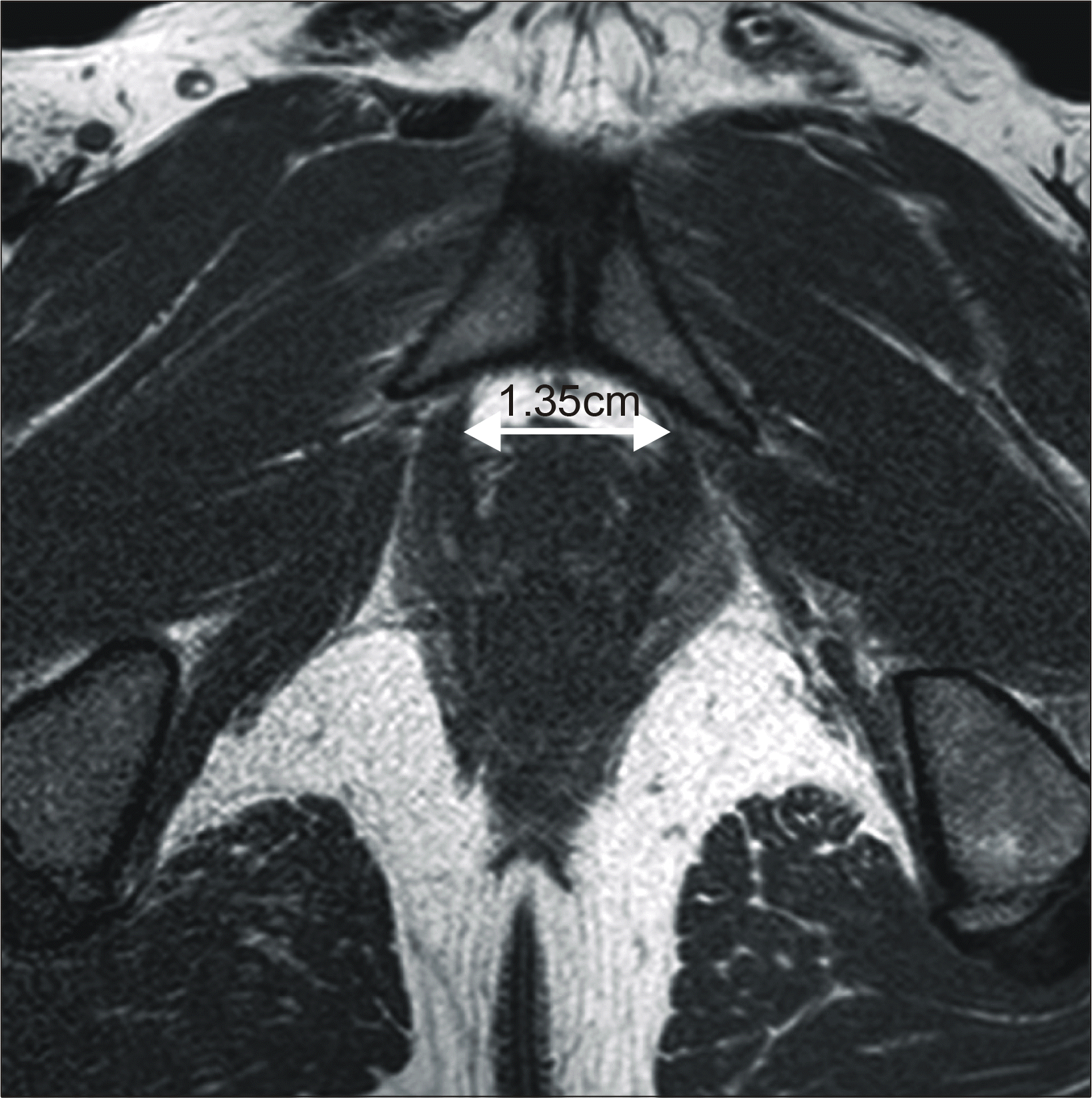 | Fig. 1.Greatest diameter of dorsal vein is measured on axial image of Tl-weighted magnetic resonance imaging. |
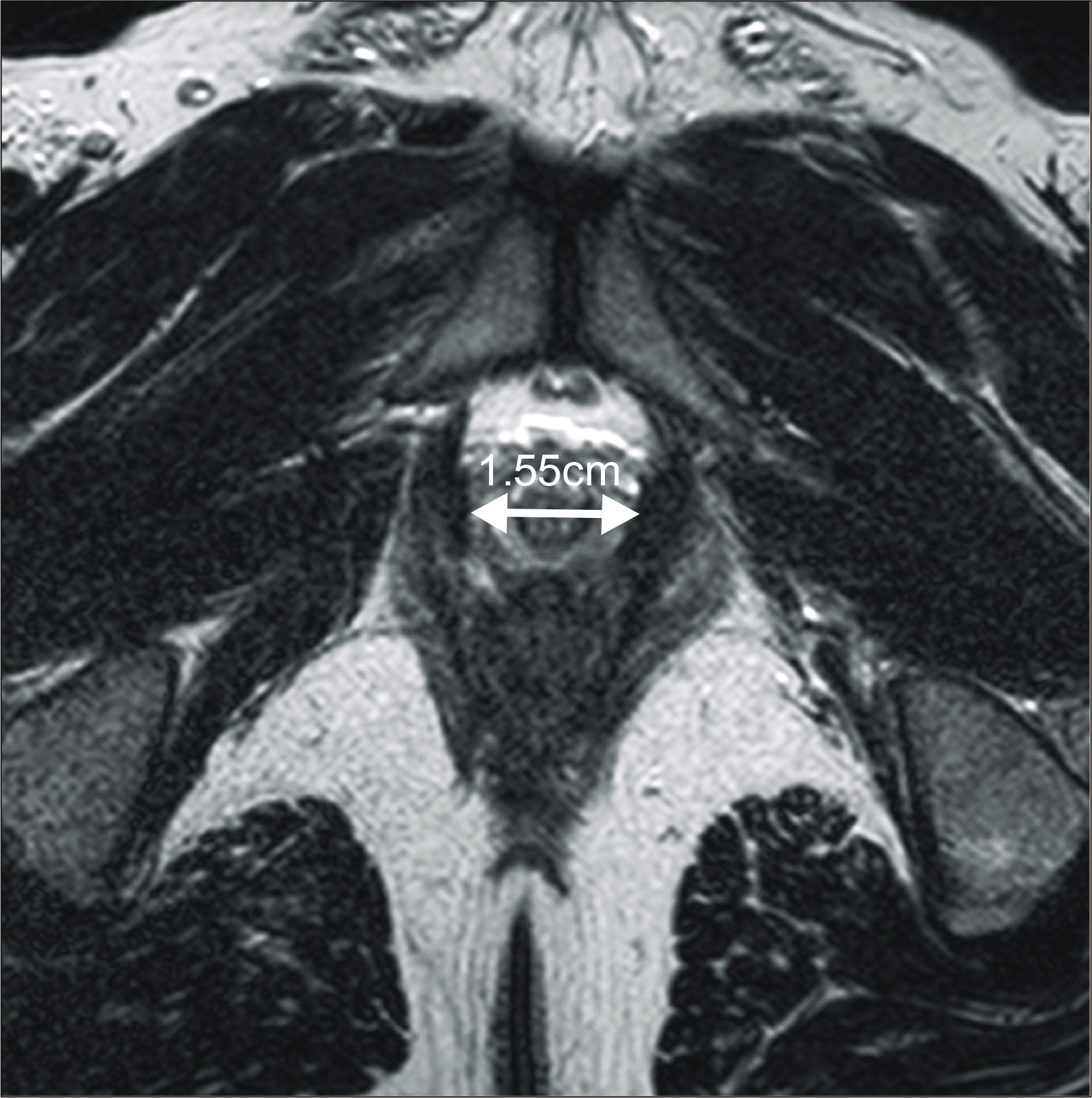 | Fig. 2.Greatest diameter of membranous urethra is measured on axial image of T2-weighted magnetic resonance imaging. |
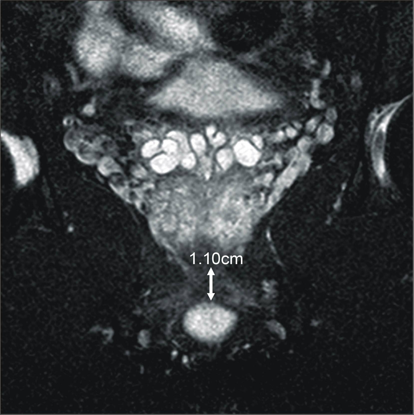 | Fig. 3.Membranous urethral length is measured on coronal image of T2-weighted magnetic resonance imaging. |
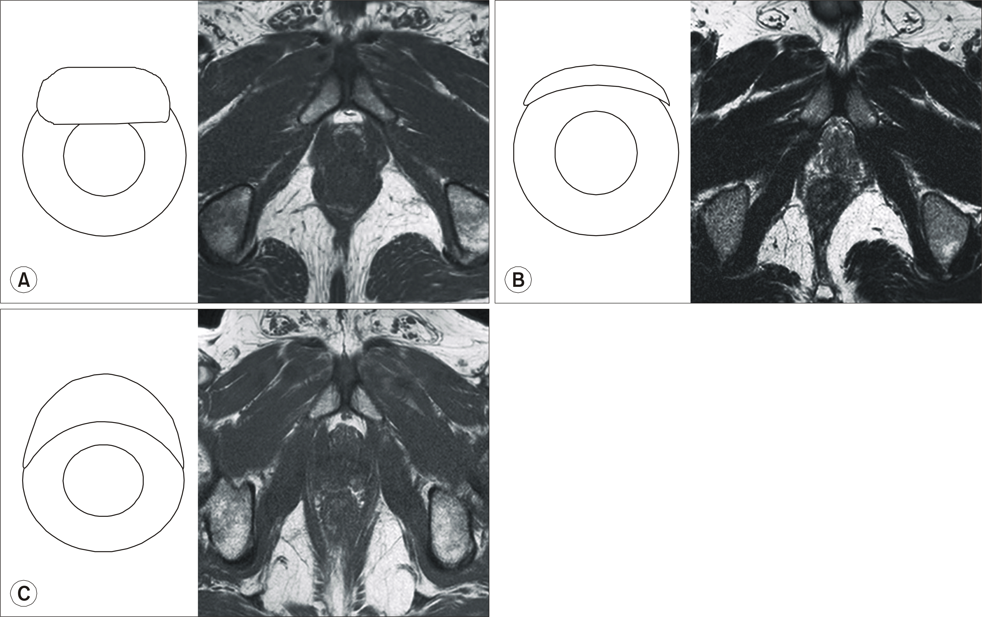 | Fig. 4.Variable shapes of dorsal vein demonstrated on axial image of magnetic resonance imaging. |
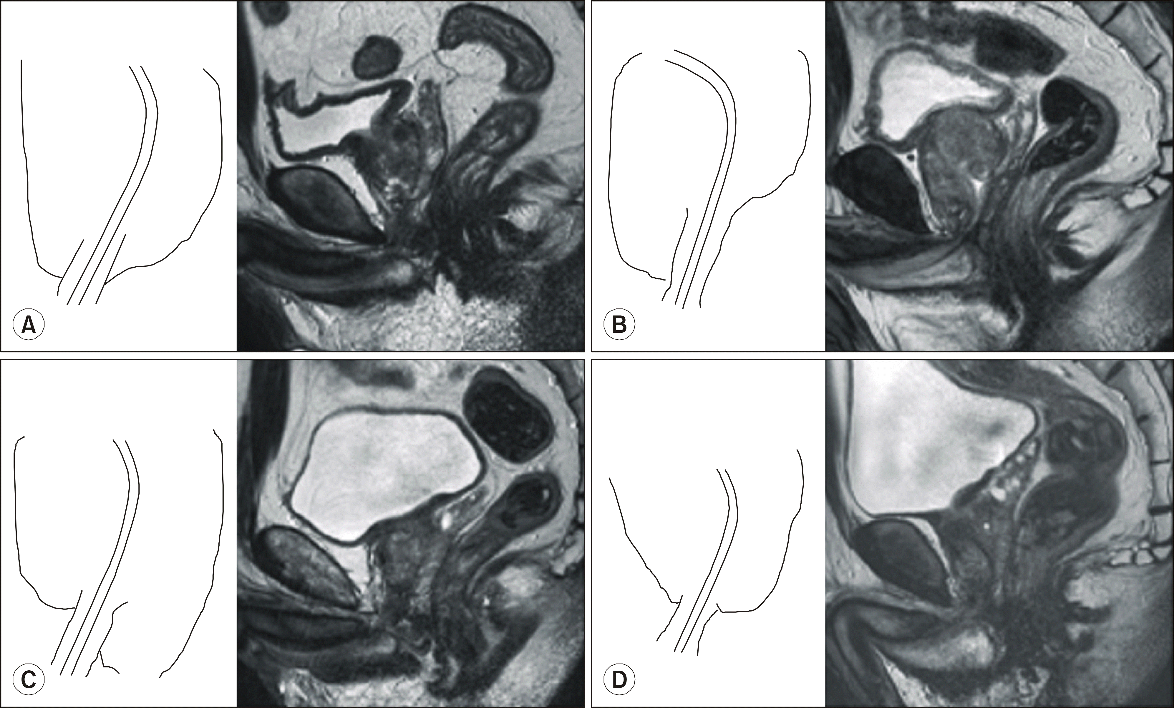 | Fig. 5.Variable shapes of prostate demonstrated on midsagittal image of magnetic resonance imaging. |
Table 1.
Patient characteristics
Table 2.
Association of various factors with blood loss during radical retropubic prostatectomy on multivariate analysis
Table 3.
Association of various factors with early recovery of continence following radical retropubic prostatectomy on multivariate analysis
 XML Download