

 PDF
PDF ePub
ePub Citation
 Print
Print


Abstract
Purpose
Increasing evidence suggests that Randalls plaque contributes to the pathogenesis of urinary stone formation. The purpose of our study is to compare the incidence of the abnormal metabolic stone risk factors between the calcium stone former with papillary calcification and the calcium stone former without papillary calcification on unenhanced spiral computed tomography (CT).
Materials and Methods
A series of patients with calcium stones (n=49) underwent unenhanced spiral CT and complete metabolic evaluation after they consumed a random diet for 1 month after stone removal. Of the 49 patients, 38 patients showed papillary calcification on unenhanced spiral CT and 11 patients did not. Their blood was evaluated by using a multichannel analysis sequential multichannel autoanalyzer (SMA)-20 and PTH tests. The 24-hour urinary constituents were assayed for calcium, oxalate, citrate, total volume, phosphorus and sodium. We compared the incidence of abnormal metabolic risk factors between the two groups. Statistical analysis was performed by chi-square tests.
Results
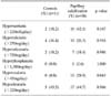
The incidences of hyperuricemia were 27.3% in the patients without papillary calcification and 31.6% in the patients with papillary calcification. The incidences of hypernatriuria were 18.2% versus 42.1%, the incidences of hypercalciuria were 36.4% versus 26.3%, the incidences of hyperuricosuria were 18.2% versus 18.4%, the incidences of hyperoxaluria were zero versus 28.9%, and the incidences of hypocitraturia were 45.5% versus 44.7%, respectively. The difference between the two groups was statistically significant only for hyperoxaluria (p=0.043).
Figures and Tables
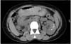 | Fig. 1Papillary calcification in the left kidney on unenhanced spiral computed tomography in our study (white arrows).
|
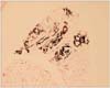 | Fig. 2von Kossa stain. This is a low magnification light microscopic image of a papillary biopsy specimen from a calcium oxalate stone patient with papillary calcification on unenhanced spiral computed tomography. The site of calcium deposits is seen in the interstitial tissue and it stained black by the von Kossa stain for calcium histochemistry (Magnification, ×100).
|
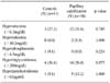
Table 1
Results of serum analysis for the patients with and without papillary calcification (mean±SD)
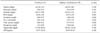
![]()
Table 2
Results of 24-hour urine analysis for the patients with and without papillary calcification (mean±SD)
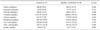
![]()
References
1. Leusmann DB, Blaschke R, Schmandt W. Results of 5,035 stone analyses: a contribution to epidemiology of urinary stone disease. Scand J Urol Nephrol. 1990. 24:205–210.
2. Westbury EJ. Some observations on the quantitative analysis of over 1000 urinary calculi. Br J Urol. 1974. 46:215–227.
3. Yoshida O, Okada Y. Epidemiology of urolithiasis in Japan: a chronological and geotraphical study. Urol Int. 1990. 45:104–111.
4. Borghi L, Meschi T, Amato F, Briganti A, Novarini A, Giannini A. Urinary volume, water and recurrences in idiopathic calcium nephrolithiasis: a 5-year randomized prospective study. J Urol. 1996. 155:839–843.
5. Cupisti A, Morelli E, Lupetti S, Meola M, Barsotti G. Low urine citrate excretion as main risk factor for recurrent calcium oxalate nephrolithiasis in males. Nephron. 1992. 61:73–76.
6. Byeon SS, Kim HH, Kim SH. Analysis of the urinary stone components using chemical analysis method. Korean J Urol. 1996. 37:179–186.
7. Uribarri J, Oh MS, Carroll HJ. The first kidney stone. Ann Intern Med. 1989. 111:1006–1009.
8. Coe FL, Parks JH. The pathogenesis and treatment of kidney stones. N Engl J Med. 1992. 327:1141–1152.
9. Smith LH. The pathophysiology and medical treatment of urolithiasis. Semin Nephrol. 1990. 10:31–52.
10. Randall A. The origin and growth of renal calculi. Ann Surg. 1937. 105:1009–1027.
11. Low RK, Stoller ML. Endoscopic mapping of renal papillae for Randall's plaques in patients with urinary stone disease. J Urol. 1997. 158:2062–2064.
12. Whalley NA, Moraes MF, Shar TG, Pretorius SS, Meyers AM. Lithogenic risk factors in the urine of black and white subjects. Br J Urol. 1998. 82:785–790.
13. Seftel A, resnick MI. Metabolic evaluation of urolithiasis. Urol Clin North Am. 1990. 17:159–169.
14. Johnson CM, Wilson DM, O'Fallon WM, Malek RS, Kurland LT. Renal stone epidemiology: a 25-years study in Rochester, Minnesota. Kidney Int. 1979. 16:624–631.
15. Carr LK, D'A Honey J, Jewett MA, Ibanez D, Ryan M, Bombardier C. New stone formation: a comparison of extracorporeal shock wave lithotripsy and percutaneous nephrolithotomy. J Urol. 1996. 155:1565–1567.
16. Sun BY, Lee YH, Jiaan BP, Chen KK, Chang LS, Chen KT. Recurrence rate and risk factors for urinary calculi after extracorporeal shock wave lithotripsy. J Urol. 1996. 156:903–905.
17. Hering LC. Observation on the analysis of ten thousand urinary calculi. J Urol. 1962. 88:545–562.
18. Francois B, Cahen R, Pascal B. Inhibitors of urinary stone formation in 40 recurrent stone formers. Br J Urol. 1986. 58:479–483.
19. Cochran M, Hofhkinson A, Zarembski PM, Anderson CK. Hyperoxaluria in adults. Br J Surg. 1968. 55:121–128.
20. Randall A. The etiology of primary renal calculus. Int Abstr Surg. 1940. 71:209–240.
21. Evan AP, Lingeman JE, Coe FL, Parks JH, Bledsoe SB, Shao Y, et al. Randall's plaque of patients with nephrolithiasis begins in basement membranes of thin loops of Henle. J Clin Invest. 2003. 111:607–616.
22. Kuo RL, Lingeman JE, Evan AP, Paterson RF, Bledsoe SB, Kim SC, et al. Endoscopic renal papillary biopsies: a tissue retrieval technique for histological studies in patients with nephrolithiasis. J Urol. 2003. 170:2186–2189.
23. Low RK, Stoller ML, Schreiber CK. Metabolic and urinary risk factors associatied with Randall's papillary plaques. J Endourol. 2000. 14:507–510.
 XML Download