

 PDF
PDF ePub
ePub Citation
 Print
Print


A pelvic kidney is frequently associated with Mullerian abnormalities such as a bicornuate or unicornuate uterus, clubfoot deformities, and cloacal exstrophy. In some cases it can appear as one component of a multi-malformation syndrome. However, a pelvic kidney with a didelphic uterus is an extremely rare finding. A didelphic uterus is usually associated with renal agenesis. Moreover, a pelvic kidney with a single ectopic ureter into the urethra is also rare. We present here a case of a huge ectopic dysplastic kidney that was associated with a didelphic uterus and a single urethral ectopic ureter.
CASE REPORT
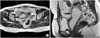
A 32-year-old woman presented with a lifelong history of lower abdominal discomfort and intermittent urinary incontinence, particularly, during the period of maternity. She had a 4-year-old son who had been delivered by an uncomplicated Caesarian section. The screening blood tests, urinalysis, and cystoscopy were normal. Vaginoscopy and colposcopy with a close inspection of the vaginal sidewall also revealed no abnormalities. Intravenous pyelogram (IVP) showed nonvisualization of the right kidney and the voiding cysturethrogram (VCUG) showed no vesicoureteral reflux. A abdominopelvic MRI showed a multicystic left kidney and a large ovoid cystic mass (5×13cm) between the two uterine bodies (didelphic uterus) (Fig. 1). This was considered to be a dysplastic right pelvic kidney with a dilated ureter running close to the urethra. The transrectal ultrasound sonography (TRUS) findings confirmed that a ectopic ureter was inserted into the urethra. The sex chromatin smears and chromosomal patterns were normal.
At transperitoneal laparoscopy, an umbilical camera port, two right-sided instrument ports, and two left-sided instrument ports were introduced. The pelvic kidney was observed just between two uterine bodies (Fig. 2A). This was removed, and the pedicle vessels, which supplied the ectopic kidney, were clipped and cut. We made an attempt to locate the actual spot of the orfice with a laparoscope by inserting it into the ureteral lumen but we could not see the orfice. Two vaginal injuries occurred during the dissection of the distal ureter. Several intracorporeal sutures were used to repair these injuries. The remnant distal ureter was treated with a purse-string suture. The kidney was removed with an endopouch through the umbilical port. The surgery time was 315 minutes, and the estimated blood loss was 460ml.
The pathologic examination confirmed an ectopic pelvic kidney with a grossly thickened ureter (Fig. 2B). The patient became currently fully continent and had no lower abdominal discomfort after operation.
DISCUSSION
A pelvic kidney occurs from an embryologic arrest at 8 weeks of gestation when the renal migration and rotation is complete. The factors that may prevent the orderly movement of the kidneys include ureteral bud maldevelopment, defective metanephric tissue that by itself fails to induce ascent, genetic abnormalities, and maternal illnesses or teratogenic causes.1,2 Twenty to 66% of females with simple renal ectopia have abnormalities in the reproductive organs such as a bicornuate or unicornuate uterus with atresia of one horn, a rudimentary or absent uterus and a proximal and/or distal vagina, and a duplication of the vagina.3 However, a pelvic kidney with a didelphic uterus is extremely rare.
In cases of a didelphic uterus, it is likely that the lesion also occurs from embryologic arrest at 8 weeks of gestation, which simultaneously affects the adjacent Mullerian and metanephic ducts.4 A didelphic uterus is often associated with ipsilateral renal agenesis or a duplicated kidney, a vaginal septum of varying degrees, and a hemivagina.5
Urinary incontinence in young women with a normal voiding pattern after successful toilet training is usually attributable to an ectopic ureter entering either the vagina or urethra. Such ureters commonly, occur in duplex systems in more than 80% of females with an ectopic ureter in which the upper-moiety ureter is ectopic.6 On the other side, a single-system ectopic ureter with congenital renal dysplasia is exceedingly rare.7
Laparoscopic treatment may work well in these cases. Challacombe et al8 reported the case of a dysplastic right pelvic kidney with a single vaginal ectopic ureter removed laparoscopically. Wang et al9 asserted that a laparoscopic upper pole heminephrectomy for an ectopic ureter was safe and feasible, and offered the patient the typical postoperative benefits of laparoscopic surgery. This case also showed successful treatment with the transperitoneal laparoscopic approach.
In laparoscopic pelvic surgery, several complications such as bladder injury, bowel injury, and ureter injury have been reported.10 However, in this case, there were some vaginal injuries due to the abnormal pelvic anatomy and severe adhesion between vagina and ectopic ureter, and these injuries were repaired by using laparoscopically intracorporeal sutures.

 XML Download