

 PDF
PDF ePub
ePub Citation
 Print
Print


Yong-Jin Kim, Hun-Jae Lee1, Tack Lee
Abstract
Purpose
The accuracy and reliability of a three-dimensional portable ultrasound bladder volume measurement system (BVMS), under two different angles (90° or 60° from cranial abdomen), for estimating the bladder volume was assessed.
Materials and Methods
Ultrasonographic studies of the bladder volume, using a newly developed portable (2.4 kg) BVMS (BioCon-500, Mcube Technology, Korea), with real bladder images, were conducted on 154 patients (29-77 years old; M:F=116:38), at angles of 90 and 60 degrees, on the abdomen 2 cm above the symphysis pubis. This ultrasound-estimated volume was compared with the immediately catheterized volume. Comparison of BVMS estimated volumes with the catheterized volumes was performed according to the angles using the Pearson correlation coefficient, intra-class correlation coefficient (ICC) concordance and fractional absolute error (FAE).
Results
Good agreement between the BVMS estimated and catheterized volumes was found for both angles (60: r=0.986, p<0.001, ICC=0.965; 90°: r=0.931, p<0.001, ICC=0.992). Although this was not significant, the linear correlation of the 60 degree estimation values seems to be higher than for those obtained at 90 degree's. Various factors, such as age, sex, body mass index (BMI) and diagnosis, showed no correlation with the difference between the catheterized and BVMS estimated bladder volumes.
Conclusions
Volume estimation using this BVMS is recommended as an alternative to catheterization for the determination of the bladder volumes both before and after voiding. The volume estimation of the transducer at 60 degrees, rather than that at 90 degrees, is recommended due to the field of view (FOV) limitation on ultrasound. However, these results demonstrate the need to standardize these procedures for volume estimations using BVMS.
Figures and Tables
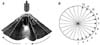 | Fig. 1Three dimensional integration process. Activating the scanning probe (A) generates 12 images, each at 15 degree angles to the adjacent image (B).
|
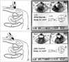 | Fig. 2The definition of ultrasonic transducer angulation and the display panel showing the actual bladder images according to the angulation. (A) 90 degree angulation. (B) Display panel of 90 degree scanning. (C) 60 Degree angulation. (D) Display panel of 60 degree scanning. Twelve images acquired in twelve planes are included in the volume determination using an integration process.
|
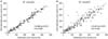 | Fig. 3Scattergram of catheter versus scan volumes, with 60 (A) and 90 (B) degrees. The two volumes are correlated at both 60 and 90 degrees (p<0.001), although the scan volume is consistent. USG: ultrasonography, ICC: intra-class correlation coefficient.
|
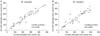 | Fig. 4Scattergram of the ultrasound estimated urine volume by two different medical staff at 60 and 90 degrees. USG: ultrasonography, ICC: intra-class correlation coefficient.
|
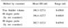
Table 1
Mean and range values of the true bladder volumes, and the ultrasound measures stratified according to examiner and method
![]()
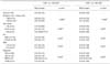
Table 2
Mean and range values of true bladder volumes, and the ultrasound measures stratified according to two different examiners

![]()
Table 3
Fractional absolute error and statistics of 154 patients according to the patients' factors
FAE: fractional absolute error, USG: ultrasonography, BMI: body mass index, NB: neurogenic bladder, SUI: stress urinary incontinence, BPH: benign prostatic hypertrophy, OAB: overactive bladder. *FAE (fractional absolute error)=| Voltrue-Volcal | / Voltrue, †calculated by Student's t-test, ‡calculated by ANOVA test
![]()
References
1. Simforoosh N, Dadkhah F, Hosseini SY, Asgari MA, Nasseri A, Safarinejad MR. Accuracy of residual urine measurement in men: comparison between real-time ultrasonography and catheterization. J Urol. 1997. 158:59–61.
2. Bih LI, Ho CC, Tsai SJ, Lai YC, Chow W. Bladder shape impact on the accuracy of ultrasonic estimation of bladder volume. Arch Phys Med Rehabil. 1998. 79:1553–1556.
3. Kim JH, Kim JH, Choi YD. The accuracy of portable ultrasound scanning in the measurement of residual urine volume. Korean J Urol. 2002. 43:933–937.
4. Lee JK, Kim SC, Nam SK. Accuracy of residual urine volume determination by ultrasonography. Korean J Urol. 1994. 35:365–369.
5. Kim JY, Park JT, Kim SC. The comparison between various formulae for calculating the residual volume by ultrasonography during volume change, and the influence of gender and age. Korean J Urol. 2004. 45:1126–1130.
6. Hwang JY, Byun SS, Oh SJ, Kim HC. Novel algorithm for improving accuracy of ultrasound measurement of residual urine volume according to bladder shape. Urology. 2004. 64:887–891.
7. Bodner DR, Witcher M, Resnick MI. Application of office ultrasound in the management of the spinal cord injury patient. J Urol. 1990. 143:969–972.
8. Alnaif B, Drutz HP. The accuracy of portable abdominal ultrasound equipment in measuring postvoid residual volume. Int Urogynecol J Pelvic Floor Dysfunct. 1999. 10:215–218.
9. Alivizatos G, Skolarikos A, Albanis S, Ferakis N, Mitropoulos D. Unreliable residual volume measurement after increased water load diuresis. Int J Urol. 2004. 11:1078–1081.
10. Marks LS, Dorey FJ, Macairan ML, Park C, deKernion JB. Three-dimensional ultrasound device for rapid determination of bladder volume. Urology. 1997. 50:341–348.
11. Yip SK, Sahota D, Chang AM. Determining the reliability of ultrasound measurements and the validity of the formulae for ultrasound estimation of postvoid residual bladder volume in postpartum women. Neurourol Urodyn. 2003. 22:255–260.
12. Hvarness H, Skjoldbye B, Jakobsen H. Urinary bladder volume measurements: comparison of three ultrasound calculation methods. Scand J Urol Nephrol. 2002. 36:177–181.
13. Hakenberg OW, Ryall RL, Langlois SL, Marshall VR. The estimation of bladder volume by sonocystography. J Urol. 1983. 130:249–251.
14. Lamonerie L, Marret E, Deleuze A, Lembert N, Dupont M, Bonnet F. Prevalence of postoperative bladder distension and urinary retention detected by ultrasound measurement. Br J Anaesth. 2004. 92:544–546.
15. Bodker B, Lose G. Postoperative urinary retention in gynecologic patients. Int Urogynecol J Pelvic Floor Dysfunct. 2003. 14:94–97.
16. Bis KG, Slovis TL. Accuracy of ultrasonic bladder volume measurement in children. Pediatr Radiol. 1990. 20:457–460.
17. Huang YH, Bih LI, Chen SL, Tsai SJ, Teng CH. The accuracy of ultrasonic estimation of bladder volume: a comparison of portable and stationary equipment. Arch Phys Med Rehabil. 2004. 85:138–141.
18. Bent AE, Nahhas DE, McLennan MT. Portable ultrasound determination of urinary residual volume. Int Urogynecol J Pelvic Floor Dysfunct. 1997. 8:200–202.
 XML Download