

 PDF
PDF ePub
ePub Citation
 Print
Print


Jinsung Park, Bumsik Hong , Taehan Park, Hyungkeun Park
, Taehan Park, Hyungkeun Park
Abstract
Purpose
The sensitivity of antegrade pyelogram (AGP), plain film radiography (KUB) and non-contrast, thin cut abdomen computerized tomography (CT) were prospectively compared for the detection of residual stones following a percutaneous nephrolithotomy.
Materials and Methods
Fifty patients (53 renal units), who had undergone a percutaneous nephrolithotomy for radiopaque renal pelvis stone, as well as a non-contrast abdomen CT 1 month postoperatively, were prospectively evaluated. The number and size of residual fragments, as determined by immediate postoperative AGP, postoperative 1 month KUB and abdomen CT, were compared.
Results
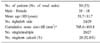
The stone-free rates according to the AGP, KUB and non-contrast CT were 73.6 (39/53), 62.3 (33/53) and 20.8% (11/53), respectively. In terms of clinically insignificant residual fragments (CIRFs), the success rates were 84.9 (45/53), 83.0 (44/53) and 41.5% (22/53), respectively. With respect to the residual stones (22 cases), which were detected by CT, but not by KUB, 45.5% (10 cases) were more than 4mm in size on CT, with a mean size of 7.4mm. The sensitivity for the detection of residual fragments was 47.6% for KUB compared to 100% for non-contrast CT. Seven patients received additional extracorporeal shock wave lithotripsy (ESWL) for residual stones following CT.
Figures and Tables
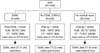
Fig. 1
Overall progress after a percutaneous nephrolithotomy in a total of 53 renal units. AGP: antegrade pyelogram, ESWL: extracorporeal shock wave lithotripsy, KUB: plain film radiography (kidney, ureter, bladder), CT: computerized tomography. *residual stone rates. CIRFs: clinically insignificant residual fragments.
References
1. Streem SB, Yost A, Mascha E. Clinical implications of clinically insignificant stone fragments after extracorporeal shock wave lithotripsy. J Urol. 1996. 155:1186–1190.
2. Khaitan A, Gupta NP, Hemal AK, Dogra PN, Seth A, Aron M. Post-ESWL, clinically insignificant residual stones: reality or myth? Urology. 2002. 59:20–24.
3. Zanetti G, Montanari E, Mandressi A, Guarneri A, Ceresoli A, Mazza L, et al. Long-term results of extracorporeal shock wave lithotripsy in renal stone treatment. J Endourol. 1991. 5:61–64.
4. Pires C, Machet F, Dahmani L, Irani J, Dore B. Sensitivity of abdominal radiography without preparation compared with computed tomography in the assessment of residual fragments after percutaneous nephrolithotomy. Prog Urol. 2003. 13:581–584.
5. Newman DM, Scott JW, Lingemen JE. Two-year follow-up of patients treated with extracorporeal shock wave lithotripsy. J Endourol. 1988. 2:163–171.
6. Graff J, Diederichs W, Schulze H. Long-term followup in 1,003 extracorporeal shock wave lithotripsy patients. J Urol. 1988. 140:479–483.
7. Beck EM, Riehle RA Jr. The fate of residual fragments after extracorporeal shock wave lithotripsy monotherapy of infection stones. J Urol. 1991. 145:6–9.
8. Lim JK, Hyun JS, Chung KH. Cost and effectiveness of different treatment options for renal calculi larger than 2cm. Korean J Urol. 2002. 43:454–458.
9. Feng MI, Tamaddon K, Mikhail A, Kaptein JS, Bellman GC. Prospective randomized study of various techniques of percutaneous nephrolithotomy. Urology. 2001. 58:345–350.
10. Lojanapiwat B. Previous open nephrolithotomy: does it affect percutaneous nephrolithotomy techniques and outcome? J Endourol. 2006. 20:17–20.
11. Lehtoranta K, Mankinen P, Taari K, Rannikko S, Lehtonen T, Salo J. Residual stones after percutaneous nephrolithotomy; sensitivities of different imaging methods in renal stone detection. Ann Chir Gynaecol. 1995. 84:43–49.
12. Pearle MS, Watamull LM, Mullican MA. Sensitivity of noncontrast helical computerized tomography and plain film radiography compared to flexible nephroscopy for detecting residual fragments after percutaneous nephrostolithotomy. J Urol. 1999. 162:23–26.
 XML Download