

 PDF
PDF ePub
ePub Citation
 Print
Print


Abstract
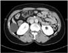
Primary mucinous cystadenomas of the retroperitoneum is extremely rare, and its histogenesis and biological behavior is unclear. Most authors suggested that it develops via mucinous metaplasia in a pre-existing mesothelium-lined cyst. We report here on a case of a 47-year-old Korean woman with primary retroperitoneal mucinous cystadenoma. Although the tumor was detected by ultrasound and computed tomography (CT), a preoperative diagnosis could not be established. The cystic tumor was successfully removed by laparoscopic surgery and microscopic examination revealed a mucinous cystadenoma. It had an ovarian stromal-like aspect and a lining of monolayer cuboidal epithelium; the tumor measured 5.0×5.0×3.5cm in size.
Figures and Tables


References
1. Min BW, Kim JM, Um JW, Lee ES, Son GS, Kim SJ, et al. The first case of primary retroperitoneal mucinous cystadenoma in Korea: a case report. Korean J Intern Med. 2004. 19:282–284.
2. Williams PP, Gall SA, Prem KA. Ectopic mucinous cystadenoma. A case report. Obstet Gynecol. 1971. 38:831–837.
3. Lauchlan SC. Metaplasias and neoplasias of Mullerian epithelium. Histopathology. 1984. 8:543–557.
4. Banerjee R, Gough J. Cystic mucinous tumours of the mesentery and retroperitoneum: report of three cases. Histopathology. 1988. 12:527–532.
5. Afriat R, Mechet I, Rachedi N, Michenet P, Bardaxoglou E, Grossetti D. Primary retroperitoneal mucinous cystadenoma: a case treated by celioscopic surgery. J Chir (Paris). 1995. 132:67–69.
 XML Download