

 PDF
PDF ePub
ePub Citation
 Print
Print


Abstract
Purpose
The purpose of this study was to report the sonographic findings of neonatal coccygeal abscess, previously not described.
Materials and Methods:
Eighteen neonates (5-18 days old) presented with swelling in the coccygeal area and by either open drainage (n=13) or follow-up after antibiotic therapy (n=5), this was diagnosed as coccygeal abscess. We retrospectively reviewed the size, shape, location, echo pattern and marginal characteristics of the abscesses, as seen on sonography, as well as their intradural content and relationship with the spine. Additional MR images (n=5) were separately reviewed.
Results:
Mean longest diameter of the abscesses was 1.5cm (range, 0.8-2.3); they were oval or round and located in the subcutaneous fat layer. Echogenicity compared with surrounding fat varied : in nine patients it was isoechoic, and in nine, hypoechoic. Internal echogenicity was homogenous in 14 patients and heterogeneous in four, and in seven cases, the margin of the abscess was well demarcated. Intradural structure and bony spines were normal, and the possibility of spinal dysraphism, could thus be excluded. All cases except one were correctly diagnosed by sonography and clinical findings å on sonography, the echogenicity of one lesion was exactly the same as that of lipoma, and it was thus misdiagnosed. In cases where sonography revealed an isoechoic mass, the use of MR excluded the possibility of lipoma. Three of five cases showed marginal or diffuse enhancement on contrast enhanced MR images.
Conclusion:
Coccygeal absesses were confined to the subcutaneous fat layer and were either iso- or hypoechoic compared with surrounding fat. In neonates, abscess formation in the coccygeal area is possible, and coccygeal abscess should therefore be included in the differentiation of coccygeal masses.
Go to : 
REFERENCES
1.Barkovich AJ. Pediatric neuroimaging. 2nd ed.New York: Raven Press;1995. 488-492.
2.Doust BD., Quiroz F., Steward JM. Ultrasonic distinction of abscesses from other intra-abdominal fluid collections. Radiology. 1977. 125:213–218.

3.Kraus R., Han BK., Babcock DS., Oestreich AE. Sonography of neck masses in children. AJR. 1986. 146:609–613.
4.Friedman AP., Haller JO., Goodman JD. Sonographic evaluation of noninflammatory neck masses in children. Radiology. 1983. 147:693–697.
5.Badami JP., Atheny PA. Sonography in the diagnosis of branchial cysts. AJR. 1981. 137:1245–1248.
6.Siegel MJ. Pediatric sonogrphy. 2nd ed.New York: Raven;1995. p. 543–547.
7.김지혜, 김인원, 연경모. 소아에서선천성척추기형의초음파진단: 자기공명영상과비교연구. 대한초음파학회지. 1992. 11:61–66.
Go to :
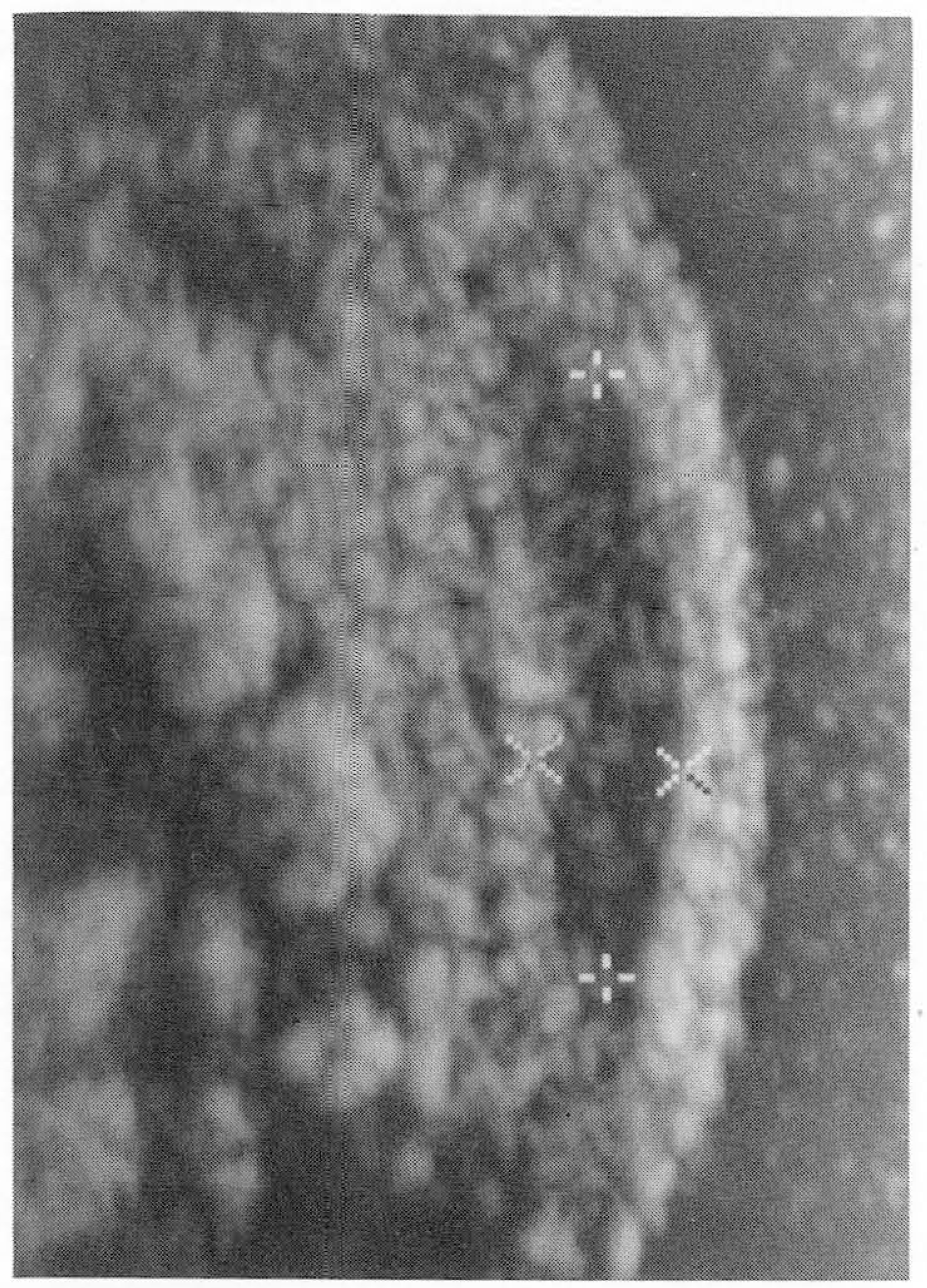
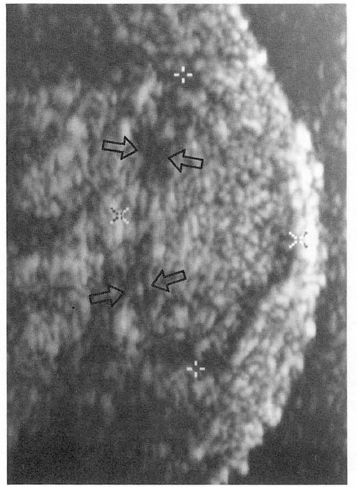 | Fig. 1.Longitudinal sonogram shows a well definded isoechoic mass (clippers) and peripheral hypoechoic rim (arrows) comparing with surrounding fat in the midline of the coccygeal subcutaneous area. |

 XML Download