Abstract
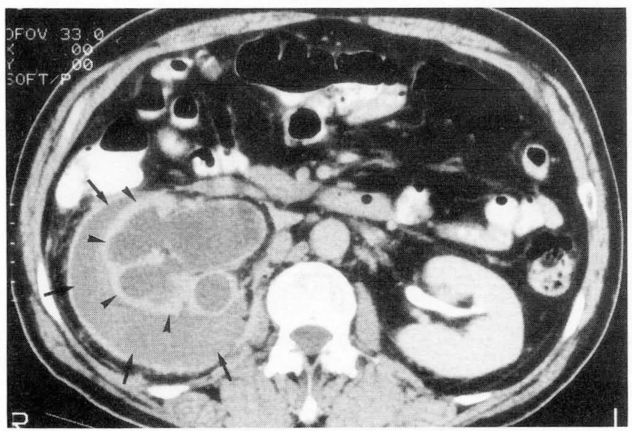
We report a unique case of spontaneous renal subcapsular hemorrhage with seeded tumor nodules along the inner surface of the renal capsule, and associated the hydronephrosis due to ureteral obstruction caused by transitional carcinoma at retero vesical junction.
Go to : 
REFERENCES
1.Errol L.., Jared J. G., Margaret L. M.: Spontaneous subcapsular and perinephric hemorrhage in end-stage kidney disease: clinical and CT findings. AJR. 1987. 148:755.
2.John S. B., Abraham M., Kevin R. L., Sab ah S.T.: Spontaneous Perinephric and subcapsular renal hemorrhage: Evaluation with CT, US, and Angiography. Radiology. 1989. 172:733.
3.Kendall A. R., Bruce A. S., Milton E. C.: Spontaneous subcapsular renal hematoma: Diagnosis and management. J Urol. 1988. 139:246.
4.Leonard M.., Melvyn K.., Paul M. S., Dunnick N.R.: Perinephric hemorrhage from metastatic carcinoma to the kidney. J Comput Assist Tomogr. 1983. 7(4):727.
5.Williams T.M.., Gardiner G.W.Spontaneous hemorrhage from osteogenic sarcoma metastasis to kidney. Can Assoc Radiol J. 1995. 46-1:51.
6.Zagoria R.J.., Dyer R.B.., Assimos E.E.., Scharling E.S.., Quinn S. F.Spontaneous perinephric hemorrhage. Invest Radiol. 1993. 28-1:81–83.
Go to : 


 PDF
PDF ePub
ePub Citation
 Print
Print


 XML Download