

 PDF
PDF ePub
ePub Citation
 Print
Print


Abstract
Purpose:
To classify perivascular change in the celiac trunk ana SMA occurring in pancreatic disease and to evaluate its significance in differential diagnosis.
Materials and Methods:
In 73 patients with pancreatic disease (42, acute pancreatitis å 14, chronic pancreatitis; 17, panreatic cancer) abdominal CT findings were retrospectively reviewed. We defined infiltration as linear or irregular density and thickeningô’ as presence of a soft tissue mantle surrounding the vessel, and statistically evaluated the usefulness of these factors for the differential diagnosis of pancreatic diseases.
Results:
In 13/42 cases of acute pancreatitis (31%), 4/l4 of chronic pancreatitis (28.6%), and 6/l7 of pancreatic cancer (35.3%), periceliac infiltration was observed å the frequencies were not statistically significant (p=0.916). Peri-SMA infiltration was demonstrated in 9/42 of acute pancreatitis 4%), 4/14 of chronic pancreatitis (28.6%), and 5/l7 of pancreatic cancer (29.4%);again, these frequencies were not statistically significant (p=0.758). Thickening of the celiac trunk and SMA was observed only in pancreatic cancer, in 3/l7 (17.6%) and 7/17(41.2%) cases, respectively, with statistical significance (P〈0.05).
Conclusion:
Thickening of the celiac trunk and SMA is a valuable flndinp in the differential diagnosis of pancreatic inflammatory disease and pancreatic cancer. When applied to the differential diagnosis of pancreatic disease, perivascular change should be classified as either infiltration or thickening.
Go to : 
REFERENCES
1.Megibow A J., Bostniak MA., Ambos MA., Beranhaum ER. Thickening of the celiac axis and/or superior mesenteric artery: A sign of pancreatic carcinoma on CT. Radiology. 1981. 14:449–453.
2.Mitchell DG., Hill MC., Cooper R, et al. The superior mesenteric artery fat plane: Is obliteration pathognomonic of pancreatic carcinoma? J Comput Assist Tomogr. 1987. 11(3):247–253.

3.Baker ME., Cohan RH., Nadel SN., Leder RA., Dunnick NR. Obliteration of the Fat Surrounding the Celiac Axis and Superior Mesenteric Artery Is Not a Specific CT Fundings of Carcinoma of the Pancreas. AJR. 1990. 155:991–994.
4.Luetmer PH., Stephens DH., Fischer AP. Obliteration of periarterial retropancreatic fat on CT in pancreatitis: an exception to the rule. AJR. 1989. 153:63–64.
5.Schulte SJ., Baron RL., Freeny PC., Patten RM., Gorell HA., Maclin ML. Root of the Superior Mesenteric Artery in Pancreatitis and Pancreatic Carcinoma: Evaluation with CT. Radiology. 1991. 180:659–662.
6.Baker ME. Pancreatic Adenocarcinoma: Are There Pathognomonic Changes in the Fat Surrounding the Superior Mesenteric Artery? Radiology. 1991. 180:613–614.
7.Gortenuti G., Cavallini G,Vantini I., Angelini G. Angiography in chronic pancreatitis and pancreatic cancer. Am J Gastroenterol. 1978. 70:620–626.
8.Megibow AJ. Pancreatic Adenocarcinoma: Designing the Examination to Evaluate the Clinical Questions. Radiology. 1992. 183:297–303.
9.Cubilla AL., Fortner J., Fitzgerald Ρ J. Lymph node involvement in carcinoma of the head of the pancreas area. Cancer. 1978. 41:880–887.
10.Balthazar EJ. CT diagnosis and staging of acute pancreatitis. Radiol Clin North Am. 1989. 27:19–37.
Go to :
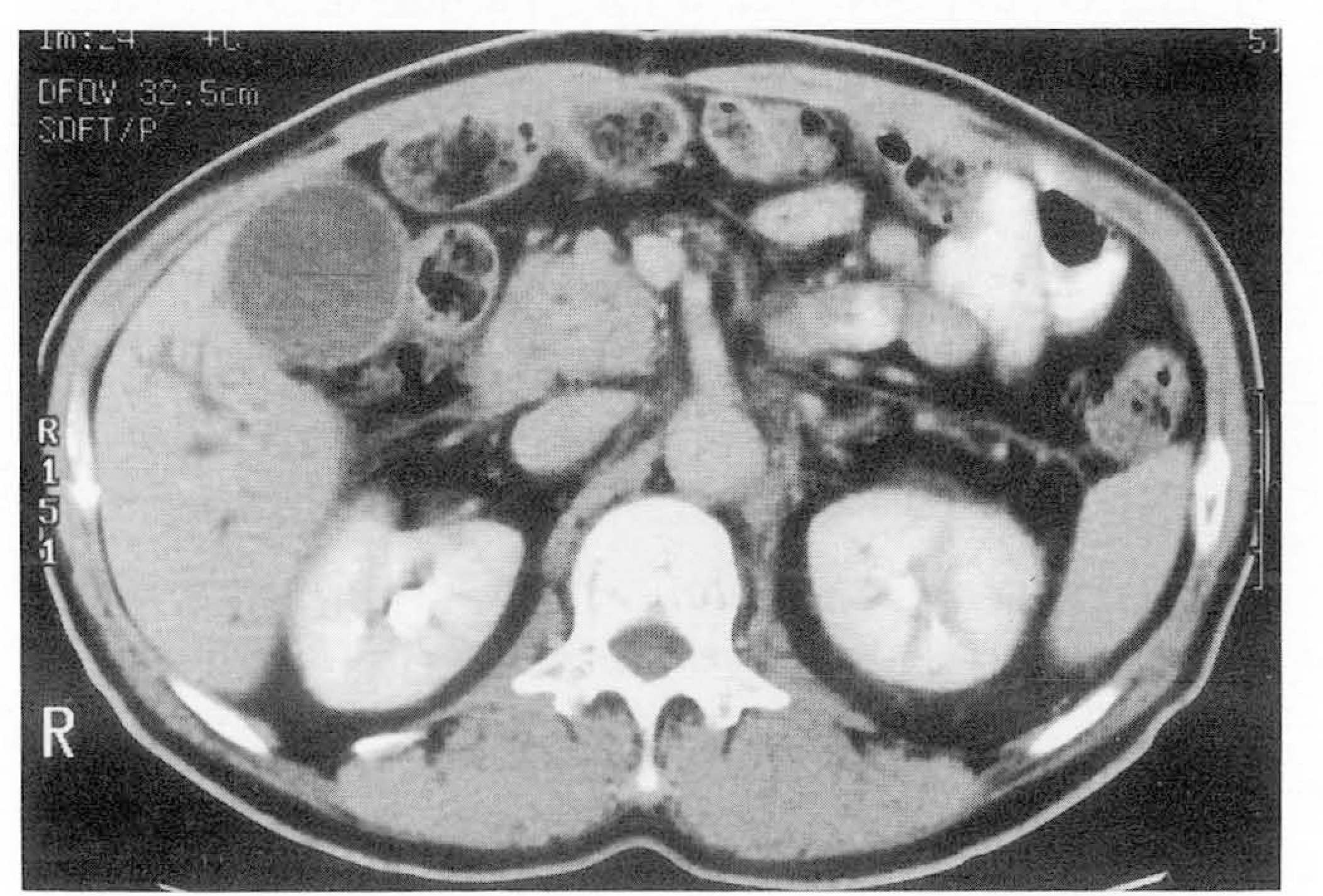
 | Fig. 1.A. Reticular infiltration of perivascular fat is demonstrated at SMA root in chronic pancreatitis 8. Perivascular fat infiltration is shown at celiac root in acute pancreatitis. |
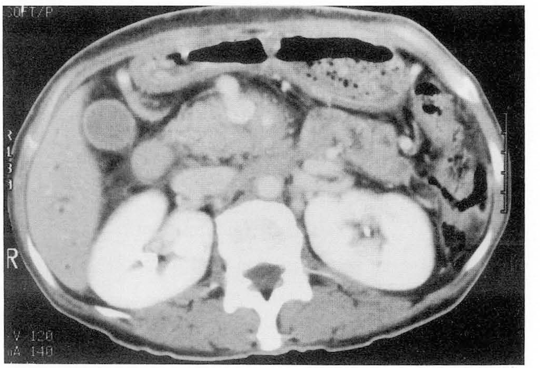
 | Fig. 2.Sixty-three year old male patient with pancreatic carcinoma å irregular infiltration is noted at SMA root area. |
 XML Download