Abstract
Trauma is the third leading cause of death, irrespective of age, and the leading cause of death in persons under 40 years of age. Most pleural, pulmonary, mediastinal, and diaphragmatic injuries are not seen on conventional chest radiographs, or are underestimated.
In patients with chest trauma, CT scanning is an effective and sensitive method of detecting thoracic injuries and provides accurate information regarding their pattern and extent.
Go to : 
REFERENCES
1.Greene R. Blunt thoracic trauma. In Greene R. eds. Syllabus: A categorical course in diagnostic radiology chest radiology. Illinois: RSNA. 1992. 297–310.
2.Toombs BD., Sandler CM., Lester RG. Computed tomography of chest trauma. Radiology. 1981. 140:733–738.

3.Kang ΕΥ., Μ ller NL. CT in blunt chest trauma: pulmonary, tracheobronchial, and diaphragmatic injuries. Semin Ultrasound CT MR. 1996. 17:114–118.

5.Mi con L., Geis L., Siderys H., Stevens L., Rodman GH. Rupture of the distal thoracic esophagus following blunt trauma: case report. J Trauma. 1990. 30:214–217.
Go to : 
 | Fig. 1.Pulmonary contusion and laceration secondary to blunt trauma. A, B. CT scan shows multifocal consolidations in left lower lobe, and ground-glass opacities are noted in both lower lobes on “lung” settings(B). C. CT scan 10 days later shows pulmonary hematoma by laceration, with clearing of pulmonary contusion. |
 | Fig. 3.Pulmonary hematoma due to blunt trauma. A. CT scan shows hematoma with air-fluid level in the posterior periphery of right lower lung. Ground-glass opacity in right lower lobe is suggestive of pulmonary hemorrhage. B. CT scan after aspiration shows marked improvement of hematoma. |
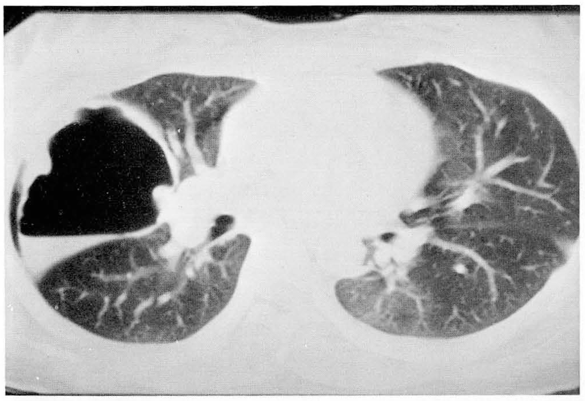
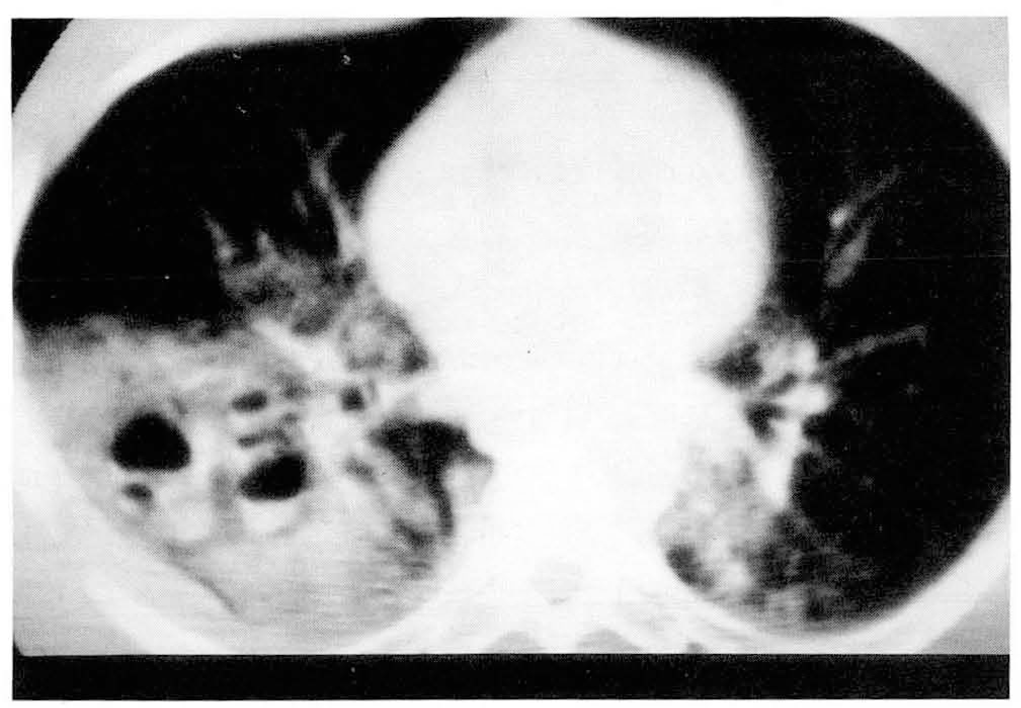 | Fig. 4.Pulmonary laceration secondary to blunt trauma. Multiple air-containing cavities with thin-walls and air-fluid levels are seen in right lung on CT scan. |
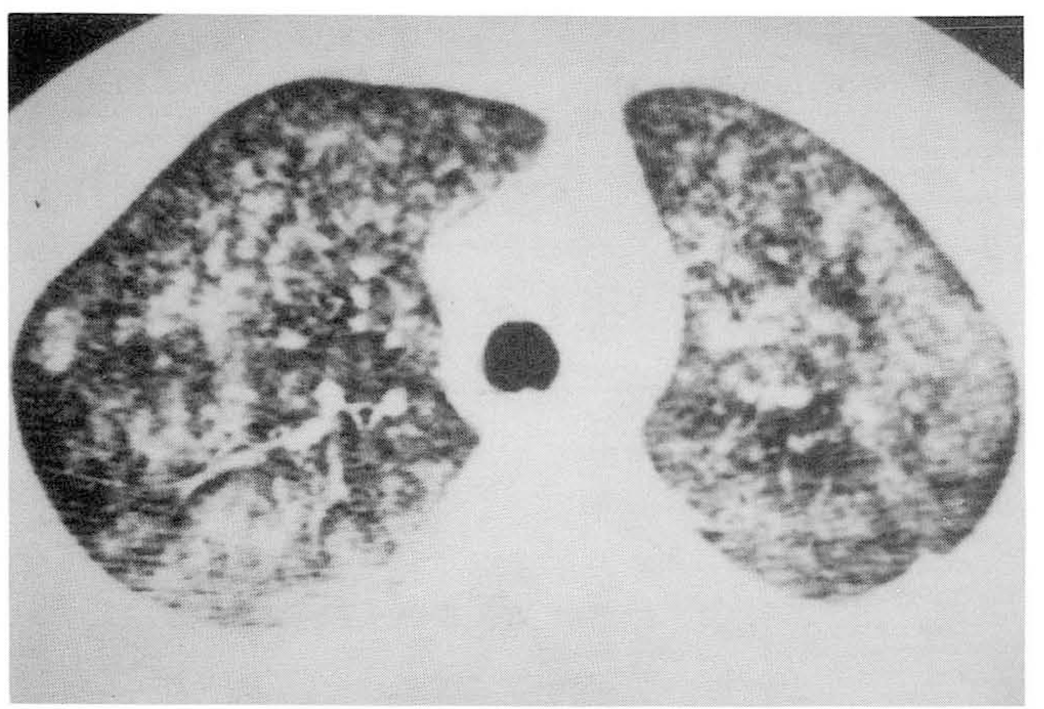 | Fig. 5.Fat embolism secondary to long bone fracture. Extensive air-space consolidation and multiple ill-defined nodules in both lungs on CT scans obtained 7 days after trauma, suggesting pulmonary hemorrhage and edema due to fat embolism. |
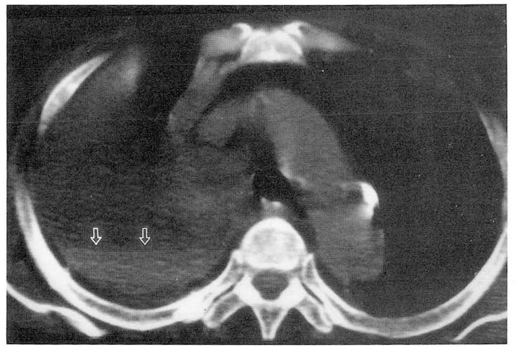 | Fig. 6.Hemothorax secondary to blunt trauma. Hemothorax with fluid-fluid level (open white arrows) is seen in the right upper hemithorax on precontrast CT. |
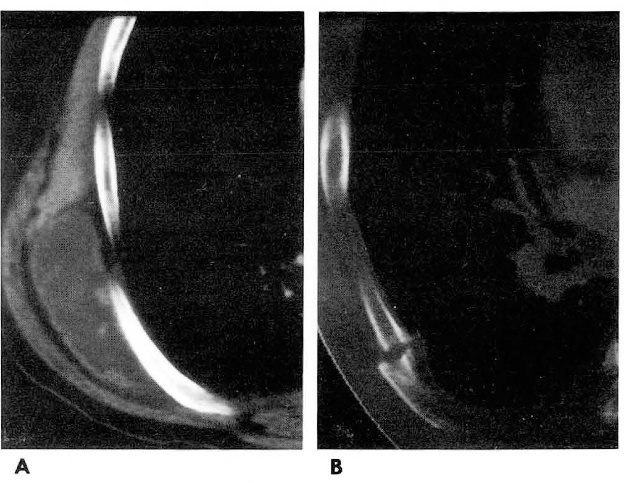
 | Fig. 7.Pneumomediastinum secondary to gun-shot injury. A, B. CT scan shows foreign body (bullet) in superior mediastinum. Ill-defined ground-glass opacities in right upper lobe (B) on CT scan with “lung settings is suggestive of pulmonary hemorrhage. |
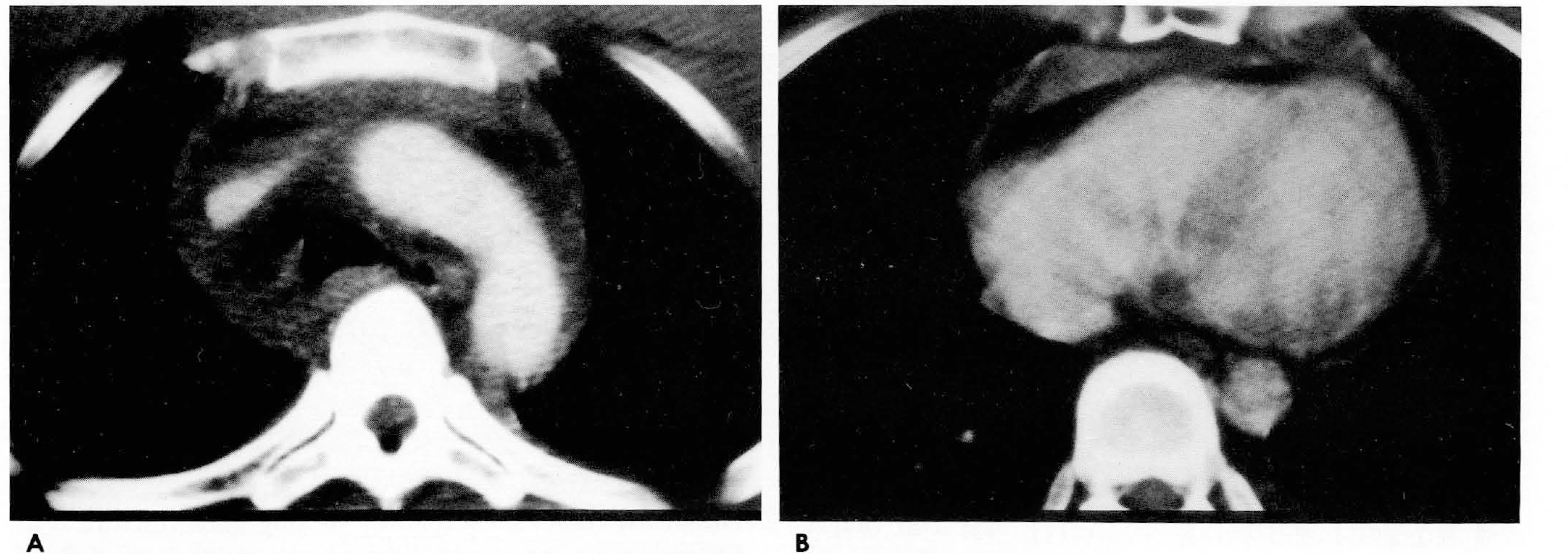 | Fig. 8.Mediastinal hemorrhage and hemopericardium secondary to blunt trauma. A. CT scan shows hemorrhage around great vessels of mediastinum. B. CT scan shows hemopericardium, which is isodense with CT attenuation of muscle in chest wall. |
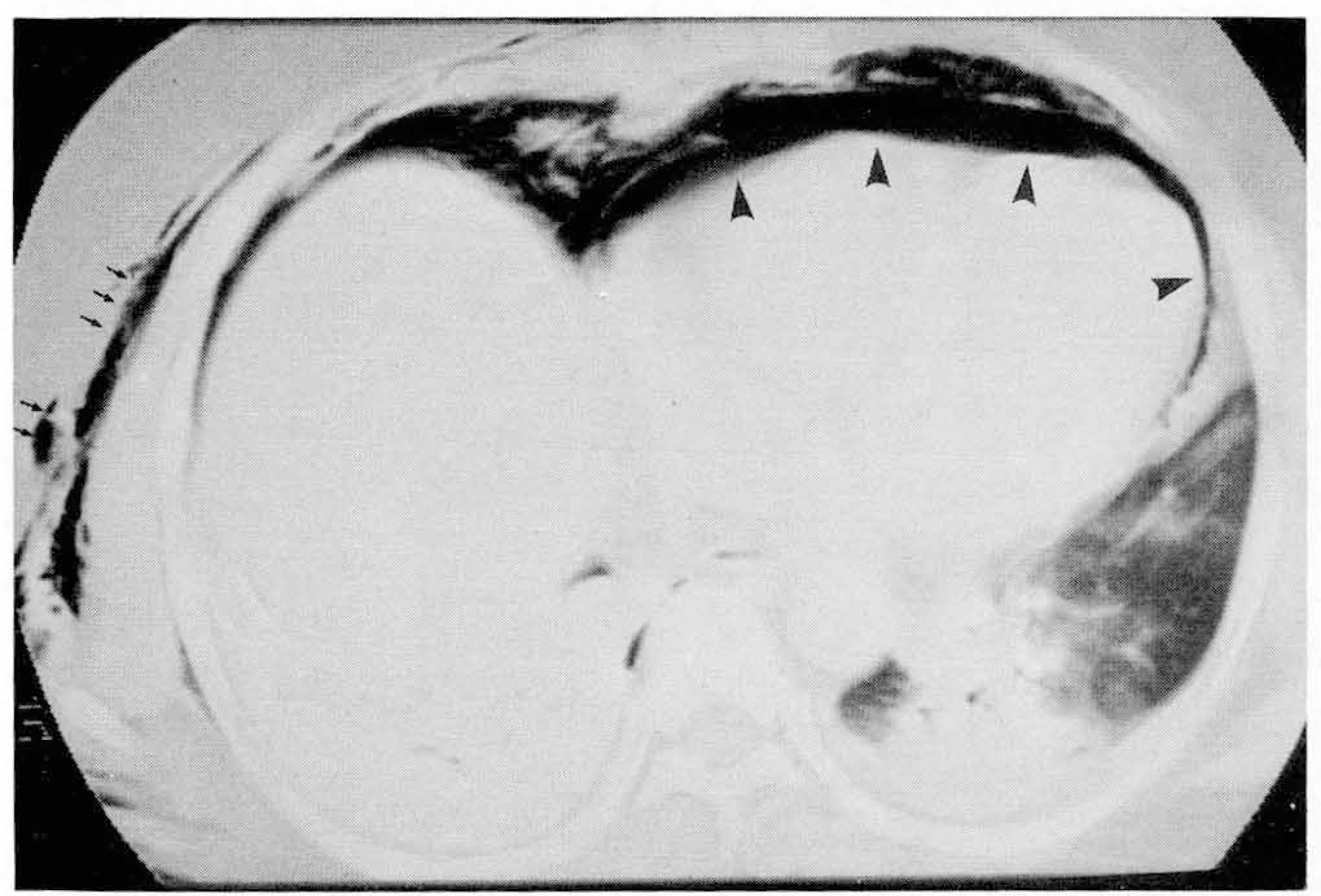 | Fig. 9.Pneumopericardium (arrowheads) and subcutaneous emphysema (small black arrows) in right chest wall on CT scan. |
 | Fig. 10.Perforation of thoracic esophagus secondary to blunt trauma. A. CT scan shows fistulous tract (black arrow) between mediastinunm and esophagus. B. CT scan shows hydropneumothorax and passive lung collapse in right lower lung. C. On esophagogram, ingested contrast media leaks from thoracic esophagus into pleural cavity. |
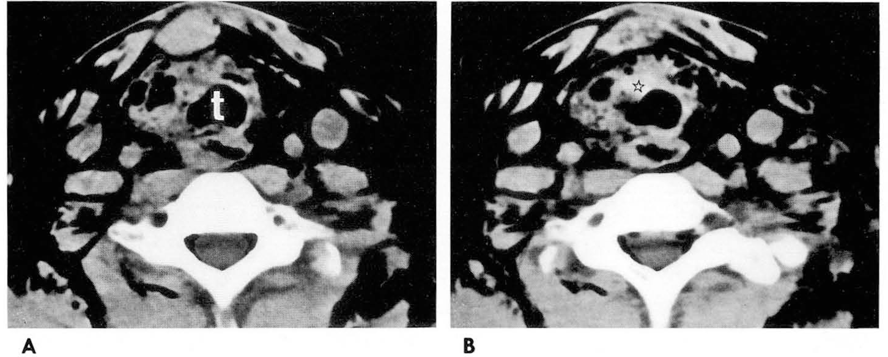 | Fig. 11.Injury of cervical trachea secondary to blunt trauma. A, B. CT scans show wall deformity of upper trachea, and irregular hematoma (open asterisk : 53 Hou- nsfield unit) and multifocal air shadows along tracheal wall, t: trachea |
 | Fig. 12.Injury of main bronchus secondary to blunt trauma. A. CT scan shows wall defect of the right main bronchus. B. Three-dimensional (3D) image shows rupture of the right main bronchus with air-pocket formation. C. 3D image after operation shows normalized right main bronchus. |
 | Fig. 13.Aortic injury secondary to blunt trauma. A, B. CT scan shows intimal flap (black arrows) along the medial aspect of aortic isthmus. Also note mediastinal hemorrhage (white arrows) lateral to aortic arch, and left hemothorax. |
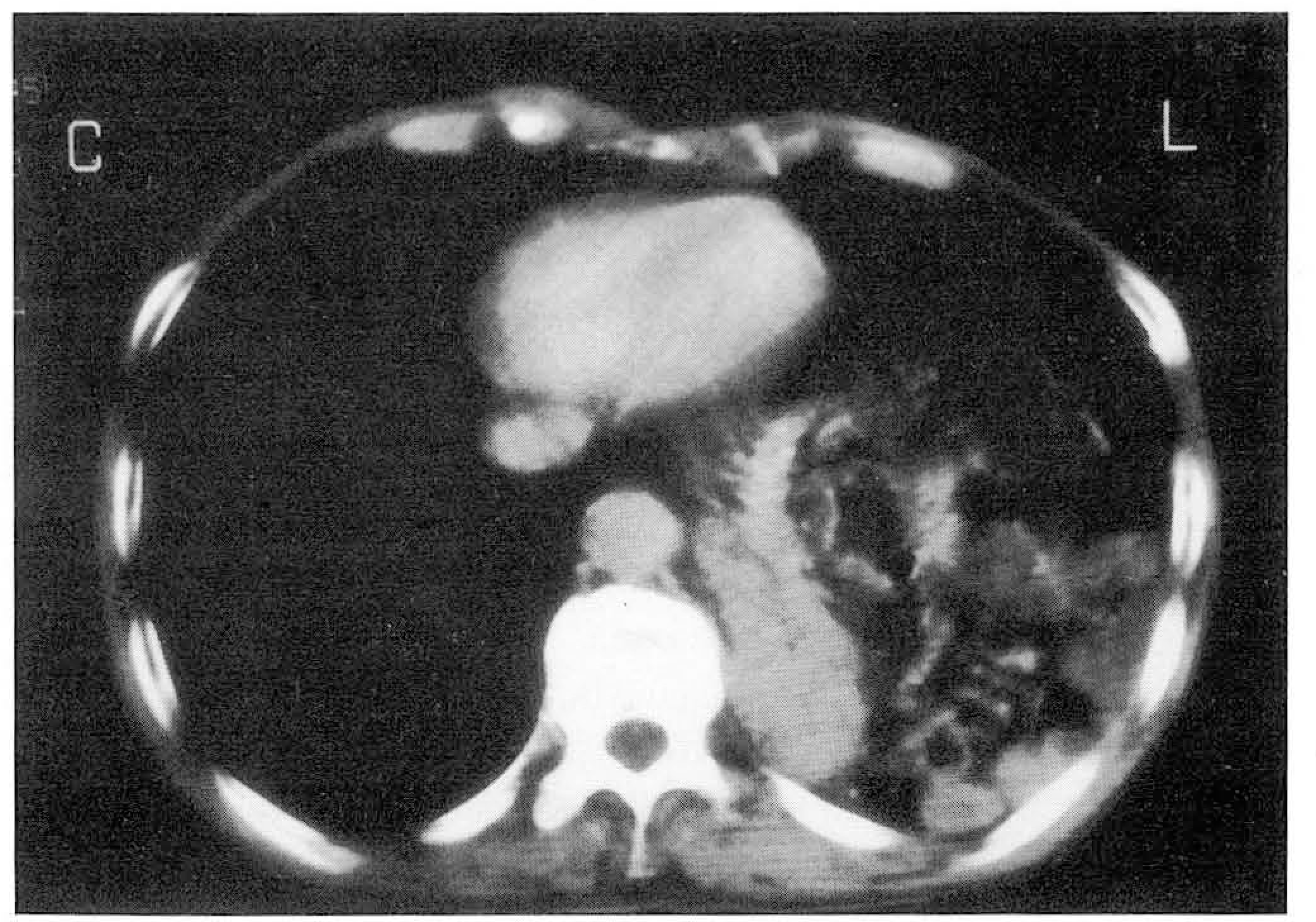
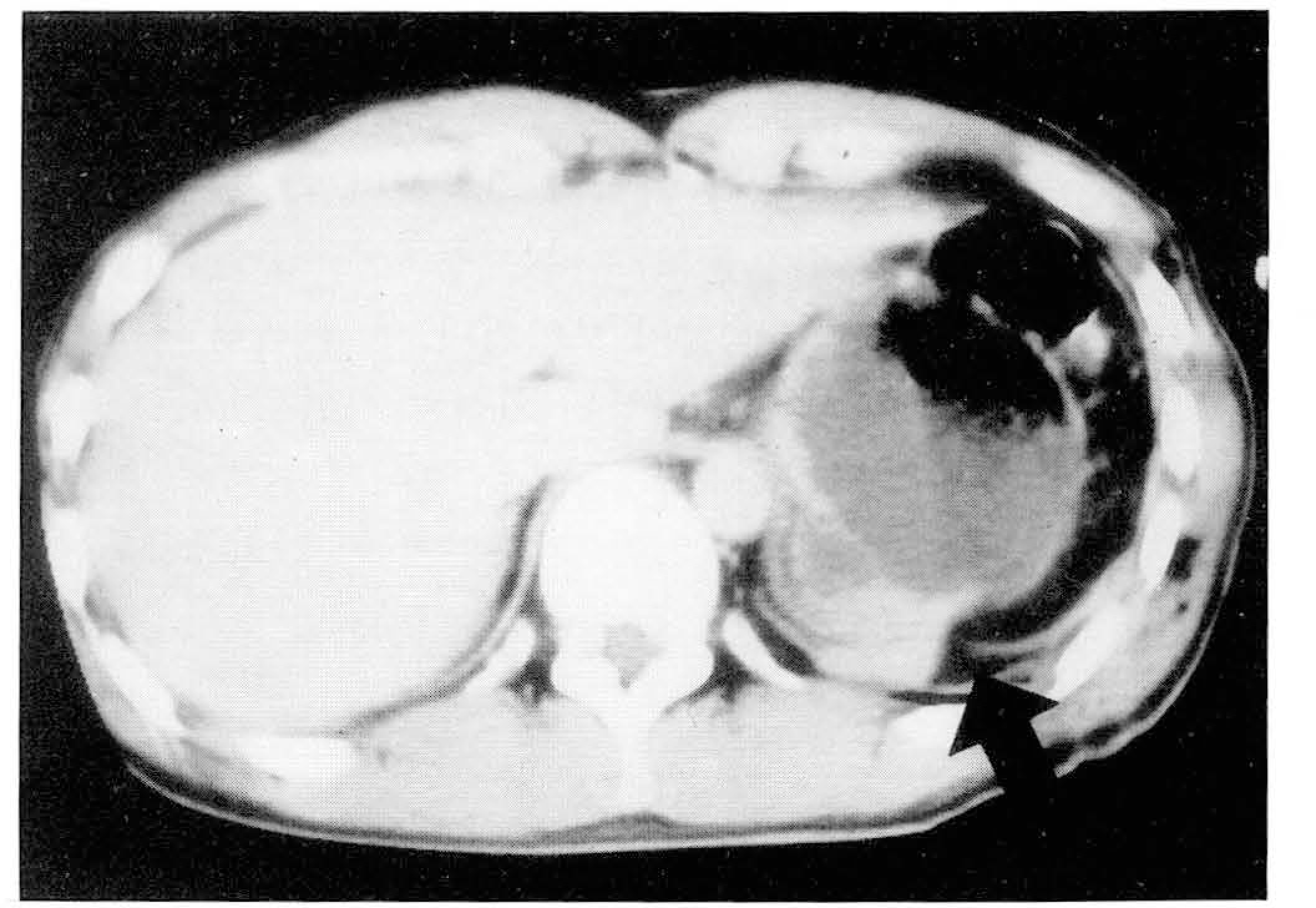
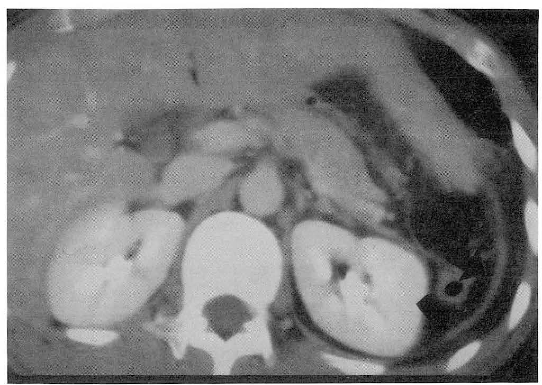 | Fig. 15.Diaphragmatic rupture secondary to blunt trauma. On CT scan at the level of upper abdomen, omental fat is seen lateral to left diaphragm (arrows). |


 PDF
PDF ePub
ePub Citation
 Print
Print


 XML Download