

 PDF
PDF ePub
ePub Citation
 Print
Print


Abstract
Purpose:
To determine the CT findings which distinguish consolidation-type bronchioloalveolar carcinoma from necrotizing pneumonia
Materials and Methods:
This study involved ten patients with pathologically-proven consolidation-type bronchioloalveolar carcinoma and 34 with necrotizing pneumonia proven pathologically either in the laboratory or clinically. We retrospectively analyzed CT features including the enhancement pattern of consolidated lung, the presence and internal density of cavity within consolidated lung, CT angiogram sign, air-bronchogram, pleural enhancement, pleural effusion, and change in extrapleural tissue and its density.
Results :
CT findings in patients with necrotizing pneumonia showed higher attenuation in marginal (94.1%) and inner (85.3%) portions in consolidated lung than in muscles(p〈 0.005); the presence of cavity(91.2%, ρ〈 0.05); cavity with fluid or air-fluid level(77.4%, ρ〈 0.005); pleural en- hancement(88.2%, ρ〈 0.00003); pleural effusion(33.3%, ρ〈0.05); and change in extrapleural tissue (64.7%, ρ {0.05). CT findings in patients with consolidation-type bronchioloalveolar carcinoma showed lower attenuation in marginal (90.0%) and inner (60.0%) portions of consolidated lung than muscles (p〈 0.005) and of cavity containing air(l00% å ρ (0.005). However, air-bronchogram and CT angiogram signs were not helpful in differentiating the two groups.
Go to : 
REFERENCES
1.Epstein DM. Bronchioloalveolar carcinoma. Semin Roentgenol. 1990. 25:105–111.

2.Hill CA. Bronchoalveolar carcinoma: a review. Radiology. 1984. 150:15–20.
3.Im JG., Han MC., Yu ΕJ, et al. Lobar bronchoalveolar carcinoma: “Angiogram Sign” on CT scan. Radiology. 1990. 176:749–753.
4.Epstein DM., Gefter WB., Miller WT. Lobar bronchioloalveolar cell carcinoma. AJR. 1982. 139:463–468.
5.Jang HJ., Lee KS., Kwon OJ., Rhee CH., Shim YM., Han JG. Bronchioloalveolar carcinoma: focal area of ground-glass attenuation at thin-Section CT as an early sign. Radiology. 1996. 199:485–488.
6.Alder B., Padley S., Miller RR., Muller NL. High-resolution CT of bronchoalveolar carcinoma. AJR. 1992. 159:275–277.
7.Wong JSL., Weisbrod GL., Chamberlain D., Herman SJ. Bronchioloalveolar carcinoma and the air bronchogram sign: a new pathologic explanation. J Thorac Imaging. 1994. 9:141–144.
8.손홍주, 김상진. 세기관지폐포암종의폐전산화단층촬영소견. 대한방사선의학희지. 1995. 32:717–723.
9.Naidich DP., Zerhouni EA., Siegelman SS. Computed tomography and magnetic resonance of the thorax. 2nd ed.New York: Raven Press;1991. ;423-427.
10.박홍석, 임정기, 유재욱, 연경모, 한만청. 괴사성폐렴: 전산화단층촬영및임상적의의. 대한방사선의학회지. 1995. 33:875–881.
11.Im JG., Webb WR., Rosen A., Gamsu G. Costal pleura: appearances at high-resolution CT. Radiology. 1989. 171:125–131.
12.Thurlbeck WM. Pathology of the lung. 2nd ed.New York: Thieme;1995. ;458-463.
13.Kuhlman JE., Fishman EK., Kuhajda FP, et al. Solitary bronchoalveolar carcinoma: CT criteria. Radiology. 1988. 167:379–382.
14.Im JG., Choi BI., Park JH, et al. CT finding of lobar bronchoalveolar carcinoma. J Comput Assist Tomogr. 1986. 10:320–322.
15.Bartlett JG., Finegold SM. Anaerobic pleuropulmonary infections. Medicine. 1972. 51:413–449.
16.Moon WK., Im JG., Yeon KM., Han MC. Complication of Klebsiella pneumonia: CT evaluation. J Comput Assist Tomogr. 1995. 19:176–181.
17.Murayama S., Onitsuka H., Murakami J., Torii Y., Masuda K., Nishihara K. “CT Angiogram Sign” in obstructive pneumonitis and pneumonia. J Comput Assist Tomogr. 1993. 17:609–612.
18.Weisbrod GL., Towers Μ J., Chamberlain DW., Herman SJ., Matzinger FRK. Thin-walled cystic lesions in bronchoalveolar carcinoma. Radiology. 1992. 185:401–405.
19.Zwirewich CV., Vefal S., Miller RR., Muller NL. Solitary pulmonary nodule: high-resolution CT and radiologic-pathologic correlation. Radiology. 1991. 179:469–476.
20.Shapiro R., Wilson GL., Yesner R., Shuman H. A useful rentgen sign in the diagnosis of localized bronchioloalveolar carcinoma. AJR. 1971. 114:516–524.
21.강은영, 이민진, 정규병. 세기관지폐포암의방사선학적소견. 대한방사선의학희지. 1992. 28:89–94.
22.김성진, 임정기, 박길선, 김학수, 한만청. Malignant vs benign pleural lesion: CT findings. 대한방사선의학회지. 1990. 26:735–742.
23.Aqunino SL., Webb WR., Gushiken BJ. Pleural exudates and transudates: diagnosis with contrast-enhanced CT. Radiology. 1994. 192:803–808.
24.Waite RJ., Carboneau RJ., Balikian JP, et al. Parietal pleural changes in empyema: apperances at CT. Radiology. 1990. 26:735–742.
Go to :
 | Fig. 1.A 64-year-old female with positive sputum culture for Pseud- omonas aeruginosa. Contrast-enhanced CT scan shows consolidation with volume expansion in the left upper lobe. Pneumonic consolidation shows higher attenuation in the marginal portion (arrowheads) than that of muscles and lower attenuation with mutiple air cavities (open arrow) in the inner portion. Note diffuse thick pleural enhancement around the consolidation(ar- rows). |
 | Fig. 2.A 67-year-old female with bronchioloalveolar cell carcinoma presenting with cough and blood tinged sputum. Contrast-enhanced CT scan shows consolidation with volume expansion in the right lower lobe. Marginal and inner portion of consolidated area have lower attenuation than that of muscles. Enhanced pulmonary vessels (open arrow) and multiple cysts (arrowhead) containing air are seen within consolidation. Note that pleural enhancement is not seen inside of the ribs(arrows). |
 | Fig. 3.A 60-year-old male with positive sputum culture for Nocardia asteroides presenting with cough and sputum. A. Contrast-enhanced CT scan shows consolidation with volume expansion in the right lower lobe. Pneumonic consolidation has higher attenuation than that of muscles. Note multiple cysts containing air (open arrow) and dilated bronchi (arrow) within consolidation. Small amount of pleural effusion is seen around consolidation. Pleural enhancement is obscured by adjacent highly enhanced consolidation. B. CT scan obtained at the lower level of (A) shows mutiple cysts with air-fluid levels (open arrow) within consolidated lung. |
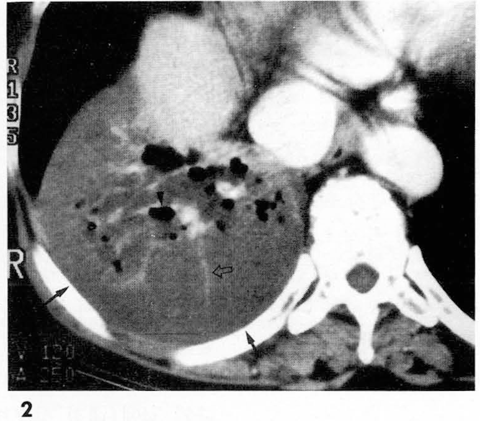
 | Fig. 4.A 55-year-old male with Klebsiella pneumonia from blood and sputum culture presenting with fever, and chill. Contrast-enhanced CT scan shows consolidation with volume expansion in the right lower lobe. Consolidated lung shows lower attenuation in the inner portion than that of muscles and equal or higher attenuation in the marginal portion(open arrow). Multiple small air cavities are seen within consolidation. Note diffuse enhancement of thickened pleura (arrowheads) and extrapleural fluid-density around consolidation (arrows). |
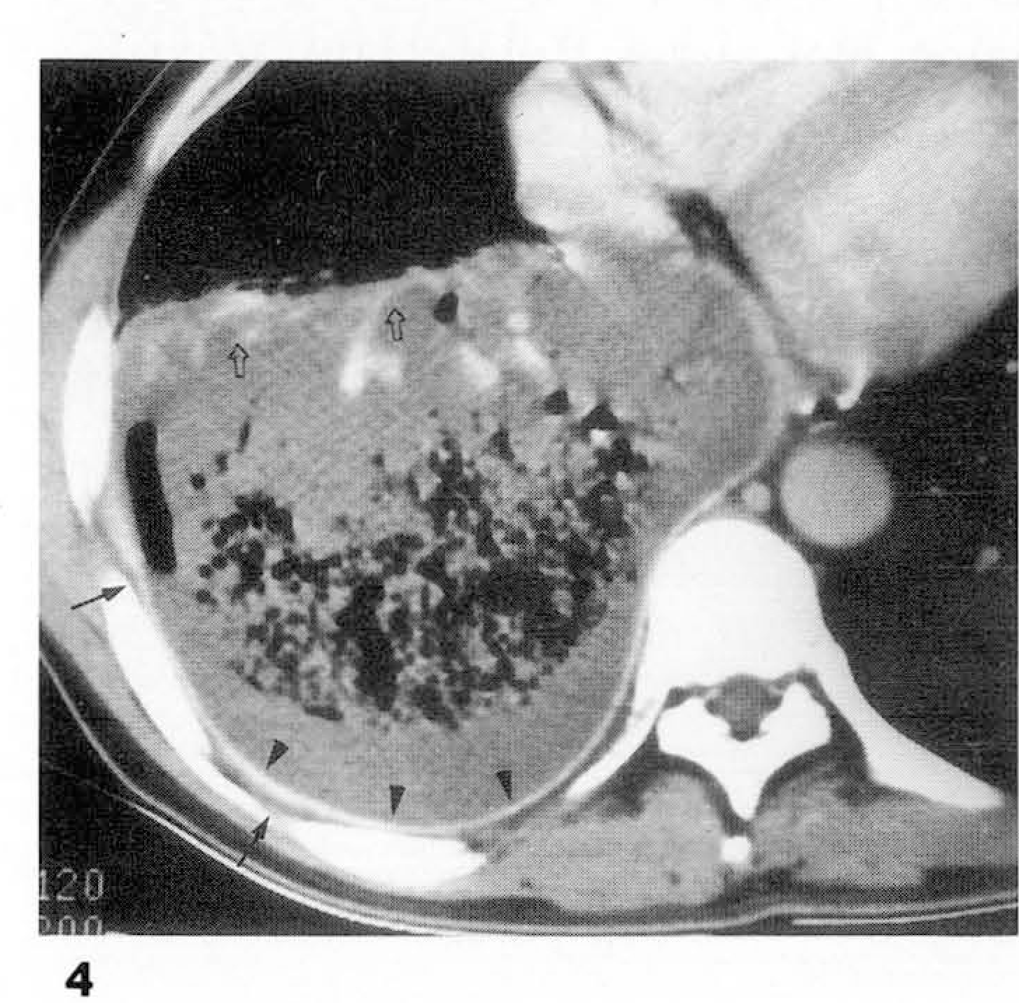
 | Fig. 5.A 48-year-old female with bronchioloalvelolar cell carcinoma in the right upper lobe and pneumonectomy state of left lung due to previous bronchioloalveolar cell carcinoma. Contrast-enhanced CT scan shows consolidation in the right upper lobe. Consolidation shows higher attenuation in the inner portion (arrowheads) than that of muscles and lower attenuation in the marginal portion. Pleural enhancement is not seen around the consolidated lung(arrow). |
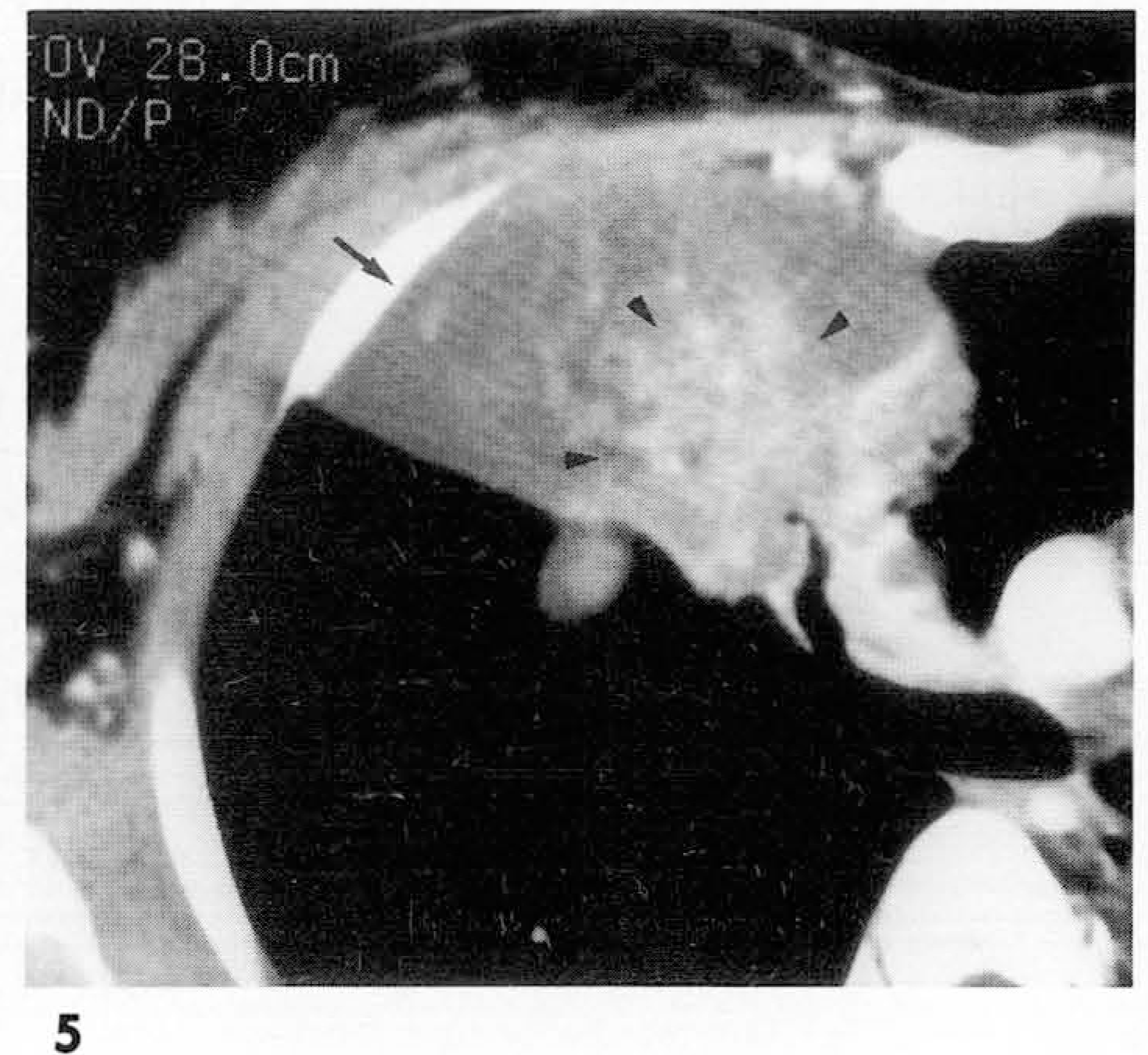
 | Fig. 6.A 53-year-old female with bronchioloalveolar cell carcinoma presenting with cough and fever. Contrast- enhanced CT scan shows low attenuating consolidation in the right lower lobe. Note that a cavities with an air densities (open arrow) within consolidation and thin enhancement of pleura (arrowheads) around the consolidation. Extrapleural fat-density (arrow) is also present. |
Table 1.
CT Attenuation of the Consolidated Lung Compared with that of Adjacent Muscles
Table 2.
CT Findings of Bronchioloalveolar Carcinoma and Necrotizing Pneumonia
 XML Download