Abstract
Purpose
To evaluate the efficacy and safety of placement of a central venous catheter with infusion port into the superior vena cava
Materials and Methods:
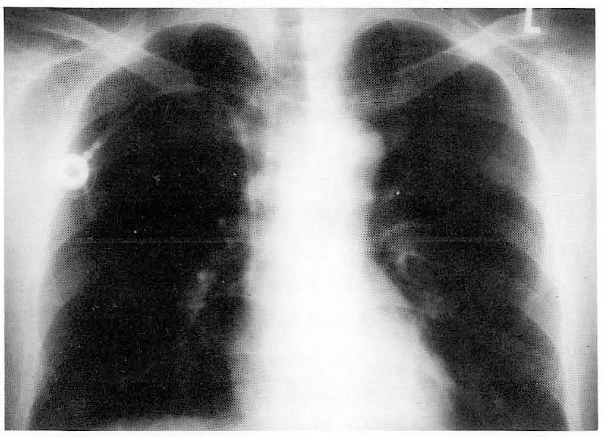
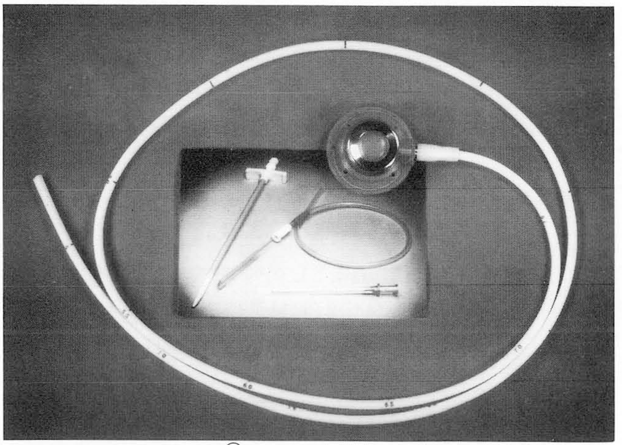
Central venous catheters with a infusion port were implanted in 21 patients (M:F = 4:17, age range: 15 — 63, mean age: 41) diagnosed as suffering from breast cancer (n=9), lymphoma(n=7), thymoma(n=2) rhabdomyosarcoma(n=2) and rectal cancer(n=l). The peripheral portion of the subclavean vein was punctured under fluoroscopic guidance during injection of contrast media at the site of the ipsilateral peripheral vein (20 cases) and under ultrasonographic guidance! 1 case). 9.6F central venous catheters placed in the superior vena cava via the subclavian vein and the connected infusion ports were implanted in the subcutaneous pocket near the puncture site of the right anterosuperior chest wall.
Results:
Catheter insertion in the superior vena cava and port implantation in the subcutaneous pocket were successful in all patients. Mean procedure time was 23 minutes and there were no early complications. Because the incision site had not healed, one patient underwent resuturing 3 weeks after the procedure. In one case, thrombotic occlusion of the catheter occurred, but successful recanalization, involving urokinase infusion, was performed. At the end of the chemotherapy schedule, at 180, 157 and 139 days after the procedure, three central venous catheters with a infusion port were removed in the radiologic suite. Catheter days are 5 days-180 days (mean, 119) from now(1997. 7. 1).
Go to : 
REFERENCES
1.Broviac JW., Cole JJ., Scribner BH. A silicone rubber atrial catheter for prolonged parenteral hyperalimentation. Surg Gynecol Obstet. 1973. 136:602–606.
2.Hickman RO., Buchner CD., Clift RA., Sanders JE., Stewart P., Thomas ED. A modified right atrial catheter for access to the venous system in marrow transplant recipient. Surg Gynecol Obstet. 1979. 148:871–875.
3.Brothers TE., von Moll LK., Niederhuber JE., Roberts JA., Walker-Andrews S., Ensminger WD. Experience with subcutaneous infusion ports in three hundred patients. Surg Gynecol Obstet. 1988. 166:295–301.
4.Damascelli B., Bonalumi MG., Terno G, et al. Interventional radiology with venous port (chemotherapy and infusion support). Eur J Radiol. 1986. 6:210–214.
5.Andrews JC., Walker-Andrews SC., Ensminger WD. Long-term central venous access with a peripherally placed subcutaneous infusion port: initial results. Radiology. 1990. 176:45–47.

6.Denny ., Jr DF. Placement and management of long-term central venous access catheters and ports. AJR. 1993. 161:385–393.

7.Aitken DR., Minton JP. The “pinch-off sign”: a warning of impending problems with permanent subclavian catheters. Am J Surg. 1984. 148:633–636.

8.Hinke DH., Zandt-Stastny DA., Goodman LR., Quebbeman ΕJ., Krzywda EA., Andris DA. Pinch-off syndrome: a complication of implantable subclavian venous access devices. Radiolgy. 1990. 177:353–356.

9.Robertson LJ., Mauro MA., Jaques PF. Radiologic placement of Hickman Catheters. Radiology. 1989. 170:1007–1009.

10.Cardella JF., Fox PS., Lawler JB. Interventional radiologic placement of peripherally inserted central catheters. J Vase Interv Radiol. 1993. 4:653–660.

11.Andrews JC., Marx MV., Williams DM., Sproat I., Walker-Andrews SC. The upper arm approach for placement of peripherally inserted central catheters for protracted venous access. AJR. 1992. 158:427–429.

12.Mauro MA., Jaques PF. Radiologic Placement of long-term central venous catheters: a review J Vase Interv Radiol. 1933. 4:127–137.
13.Lamers JS., Post PJM., Zonderland HM., Gerritsen PG., Kappers-Klunne MC., Sch tte HE. Percutaneous placement of Hickman catheters: Comparason of sonographically guided and blind techniques. AJR. 1990. 155:1097–1099.
14.Hahn ST., Pfammatter T., Cho KJ. Carbon dioxide gas as a venous contrast agent to guide upper-arm insertion of central venous catheters. Cardiovasc Intervent Radiol. 1995. 18:146–149.

15.Hovsepian DM., Bonn J., Eschelman DJ. Techniques for peripheral insertion of central venous catheters. J Vase Interv Radiol. 1993. 4:795–803.

16.Clarke DE., Raffin TA. Infectious Complications of indwelling long-term central venous catheters. Chest. 1990. 97:966–972.

17.Press OW., Ramsey PG., Larson EB., Fefer A. Hickman catheter infections in patients with malignancies. MedicinefBaltimore). 1984. 63:189–200.

18.Early TF., Gregory RT., Wheeler JR, et al. Increased infection rate in double lumen versus single lumen Hickman Catheters in Cancer Patients. South Med J. 1990. 83:34–36.
19.Moss JF., Wagman LD., Riihimaki DU, et al. Central venous thrombosis related to the silastic Hickman-Broviac catheters in an oncologic population. J Par enter Enteral Nutr. 1989. 13:397400.
20.Gray WJ., Bell WR. Fibrinolytic agents in the treatment of thrombotic disorders. Semin Oncol. 1990. 17:228–237.
21.Bern MM., Lokich JJ., Wallach SR, et al. Very low doses of warfarin can prevent thrombosis in central venous catheters: a randomized prospective trial. Ann Intern Med. 1990. 112:423428.
Go to : 


 PDF
PDF ePub
ePub Citation
 Print
Print


 XML Download