Abstract
Materials and Methods:
In eighteen patients with pathologically-proven non-Hodgkin's lymphoma in the sinonasal cavity, CT and MR images were retrospectively reviewed. CT and MR findings were analyzed for tumor location, degree of infiltration into the adjacent structure, degree of enhancement, and the presence of bone change. Tthe last-named was classified as one of four types: complete destruction, segmental destruction, thinning, or sclerotic change.
Results:
Masses in the nasal cavity (N二 17) and ethomoid sinus (N=16) were most common, and the remainder were accounted for by maxillary sinus(N=o), sphenoid sinus(N=2), and frontal sinus (N=2)· In 16 cases, the involvement of more than two sinonasal compartments was demonstrated å the deensity of these masses was shown by precontrast CT to be similar to that of facial muscles å affer contrast enhancement, all except one (15/16) showed homogeneous enhancement.
Tumor innltration of the adjacent structure was identified in the nasopharynx(N=9), anterior buccal space(N 二7), orbit(N==6), subcutaneous layer of the cheek(N=3), and infratemporal fossa (N二 3). Direct extension of the tumor from the nasal fossa to the nasopharynx or anterior buccal space was demonstrated.
Among 18 cases, bone change was seen in 12, segmental destruction in eight, comolete destruction in six, thinning in two, and sclerotic change in two. Four of the six cases with complete bone destruction showed hyperaense linear density within the mass å CT showed that after treatment, bony regrowth had occurred.
In two cases, MRI showed intermediate signal intensity of the masses on T1WI, iso or slightly high signal intensity on T2WI, and moderate enhancement on postcontrast T1WI.
Conclusion:
On CT, sinonasal lymphoma usually showed homogenous enhancement, extensively infiltration of the adjacent structure, but no massive bone destruction. Hyperdense linear density, suggesting ghost bone and seen in spite of massive bone destruction, may be a characteristic finding of sinonasal lymphoma.
Go to : 
REFERENCES
1.Freeman C., Berg JW., Cutler SJ. Occurence and prognosis of extranodal lymphoma. Cancer. 1972. 29:252–260.
2.Frierson HF., Mills SE., Innes DJ. Non-Hodgkin's lymphomas of the sinonasal region: Histologic subtypes and their clinicopathologic features. Am J Clin Pathol. 1984. 81:721–727.

3.노태연, 백호길, 원종부등. 비강비호즈킨림프종의전산화단층촬영소견. 대한방사선의학회지. 1997. 36:741–746.
4.Matsumoto S., Shi buy a Η., Tatera S., Yamazaki E., Suzuki S. Comparison of CT findings in non-Hodgkin lymphoma and squamous cell carcinoma of the maxillary sinus. Acta Radiol. 1992. 33:523–527.

5.Abbondanzo SL., Wenig BM. Non-Hodgkin's lymphoma of the sinonasal tract. Cancer. 1995. 75:1281–1291.
6.Fellbarum C., Hansmann ML., Lennert K. Malignant lymphomas of the nasal cavity and paranasal sinuses. Virchows Arch A Pathol Anat Histopathol. 1989. 414:399–405.
7.William HW., Stephen GH., Peter MB. Lymphoma of the nose and paranasal sinuses. Arch Otolaryngol. 1983. 109:310–312.
8.이경훈, 김형진, 박지현, 안인옥, 정성훈. 조영증강CT상저밀도음영을보이는비호지킨임파종: 병리조직학적유형과의관계. 대한방사선의학희지. 1994. 31:1191–1194.
9.Lodwick GS. The bones and joints. Chicago: Year book medical publishers. 1971. 361–385.
10.Agustin A., Leticia R., Renal do G., Alejandra T., Edna LG., Jose CD. Angiocentric T-cell lymphoma of the nose, paranasal sinuses and hard palate. Hematol oncol. 1992. 10:141–147.
11.Som PM., Curtin HD. Head and neck imaging. 3rd ed.St. Louis: Mosby-Year Book;1996. p. 213–215.
Go to : 
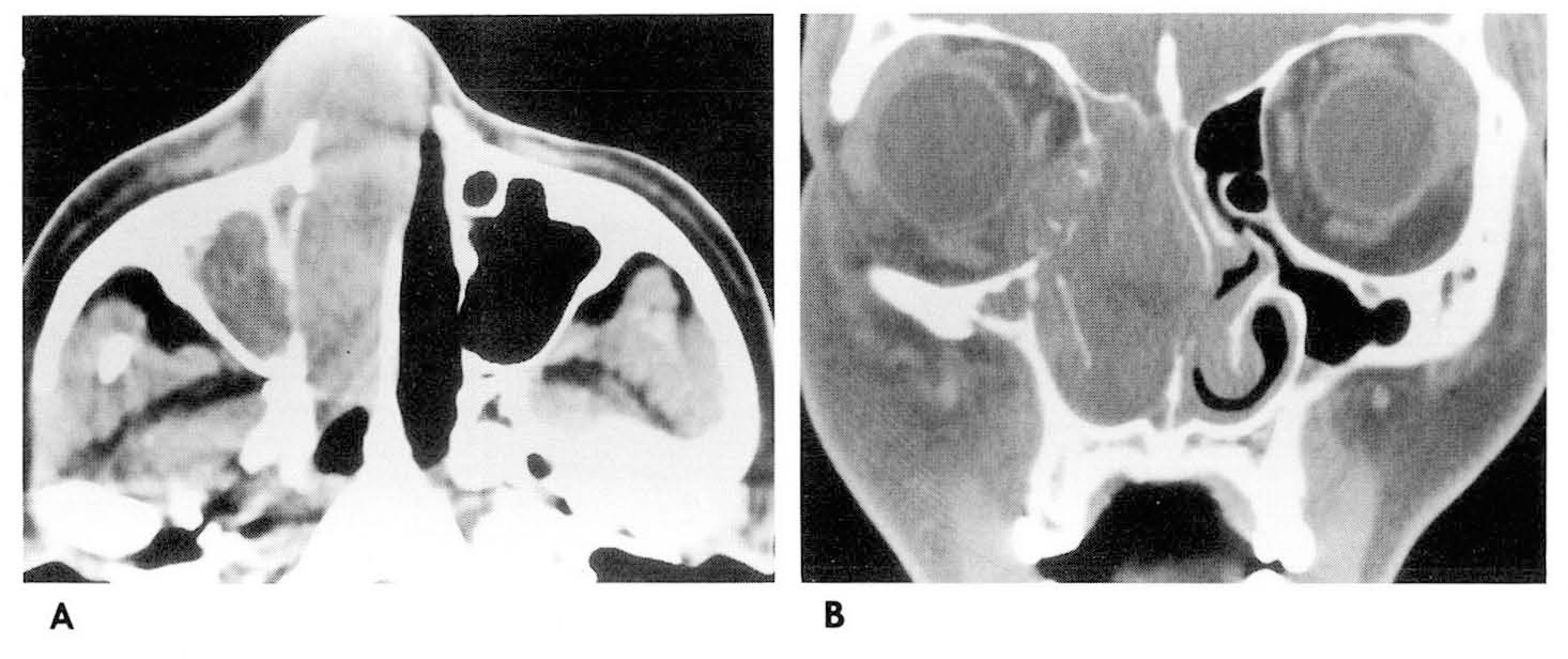
 | Fig. 1.30-year old woman with diffuse large cell type non-Hodgkin lymphoma A. Contrast-enhanced axial CT scan shows enhancing soft tissue mass of the right nasal cavity infiltrating into anterior buccal space. B. Contrast-enhanced coronal CT scan shows mass of the ethmoid sinus and nasal cavity infiltrating into the orbit through the lamina papyracea with segmental destruction. Note thinning of inferior turbinate. |
 | Fig. 2.35-year old man with diffuse mixed cell type non-Hodgkin lymphoma A. Contrast-enhanced axial scan shows homogenously enhancing mass of maxillary sinus, infiltrating into the subcutaneous layer of the cheek, the infratemporal fossa and the nasal cavity with complete bone destruction(arrow). B. Contrast-enhanced coronal scan shows a soft tissue mass in the maxillary sinus extending to the ethmoid and the sphenoid sinuses and the nasal cavity with extensive bone destruction of the maxillary sinus. Note hyperdense linear density, suggesting ghost bone(arrow). C. Follow-up CT after treatment shows the decreased size of the mass and reconstruction of the destroyed bone(arrow). |
 | Fig. 3.3-year old man with lymphoblastic type non-Hodgkin lymphoma A. Contrast-enhanced axial CT scan at the level of maxillary sinus shows enhancing soft tissue mass in the nasal cavity extending to the right infratemporal fossa with widened sphenopalatine fossa(arrow). B. Contrast-enhanced axial CT scan at the level of ethmoid sinus shows expansile mass of the ethmoid sinus, invading the both orbit with complete destruction of lamina papyracea. C. Contrast-enhanced coronal CT scan shows soft tissue mass of the nasal cavity and ethmoid sinus, infiltrating into the both orbit and the floor of the anterior cranial fossa. |
 | Fig. 4.58-year old man with large cell immunoblastic type non-Hodgkin lymphoma A. Contrast-enhanced axial CT scan shows homogeneously enhancing mass of the left ethmoid sinus. B. Tl-weighted MR image shows intermediate signal intensity tumor in the left ethmoid sinus. On the T2-weighted MR image(not shown), mass reveals slightly high signal intensity. C. Contrast-enhanced Tl-weighted MR image in the axial plane shows moderate enhancing mass. Note good differentiation of small sized sinusitis from mass(arrow). |
Table 1.
Characteristics of 18 Patients with Non-Hodgkin Lymphoma
N=nasal cavity, M=maxillary sinus, E=ethmoid sinus, F=frontal sinus, S=sphenoid sinus, A=angiocentric type N∗== nasopharynx, Β=anterior buccal space, 0=orbit, C=subcutaneous layer of the cheek, I=infratemporal fossa C∗=complete destruction, S=segmental destruction, Τ=thinning, Sc=sclerotic change, H=hyperdense linear density F/u=follow-up


 PDF
PDF ePub
ePub Citation
 Print
Print


 XML Download