Journal List > J Korean Radiol Soc > v.38(3) > 1068135

Lee, Byun, and Kim: Intracranial Angiolipoma : A Case Report1

Abstract

Intracranial angiolipoma is extremely rare. We report the radiologic findings of angiolipoma in the right middle cranial fossa, extending medially into the suprasellar and cavernous sinus region, in a 63-year-ola woman. The lesion was a relatively well marginated extra-axial tumor that showed low density on precontrast CT and marked enhancement after contrast infusion. MR imaging showed heterogeneous low signal on T1W1 mixed with hyperintense foci on T2W1, and marked enhancement after gadolinium infusion. On cerebral angiography, displacement of the right internal carotid artery by the tumor was seen. On an arteriogram of the right external carotid artery, the mass showed persistent capillary blush.

REFERENCES

1.Bowen JT. Multiple subcutaneous haemangiomas together with multiple lipomas, occurring in enormous numbers in an otherwise healthy, muscular subject. Am J Med Sci. 1912. 144:189–192.
2.Howard WR., Helwing EB. Angiolipoma. Arch Dermatol. 1960. 82:924–931.
crossref
3.Takeuchi J., Handa H., Keyaki A, et al. Intracranial Angiolipoma. Surg Neurol. 1981. 15:110–113.
crossref
4.Wilkins PR., Hoddinott C., Hourhan MD, et al. Intracranial Angilipoma. J Neurol Neurosurg Psychiatry. 1987. 50:10571059.
5.Shuangshoti S., Vajragupta L. Angiolipoma of thalamus presenting with abrupt onset suggestive of cerebrovascular disease. Clin Neuropathol. 1995. 14:82–85.
6.Prabu SS., O'Donovan DG., Gurusinghe NT. Intracranial Angiolipoma: report of two cases. Br J Neurosurg. 1995. 9:793–797.
crossref
7.Lin J J., Lin F. Two entities in angiolipoma. Cancer. 1974. 34:720–727.
8.Salibi SS., Nauta HJW., Brem H, et al. Lipomeningioma: Report of three cases and review of the literature. Neurosurgery. 1989. 25:122–126.
crossref
9.Leroux P., Hope A., Lofton S, et al. Lipomatous meningioma-An uncommon tumor with distinct radiographic findings. Surg Neurol. 1989. 32:360–365.
crossref

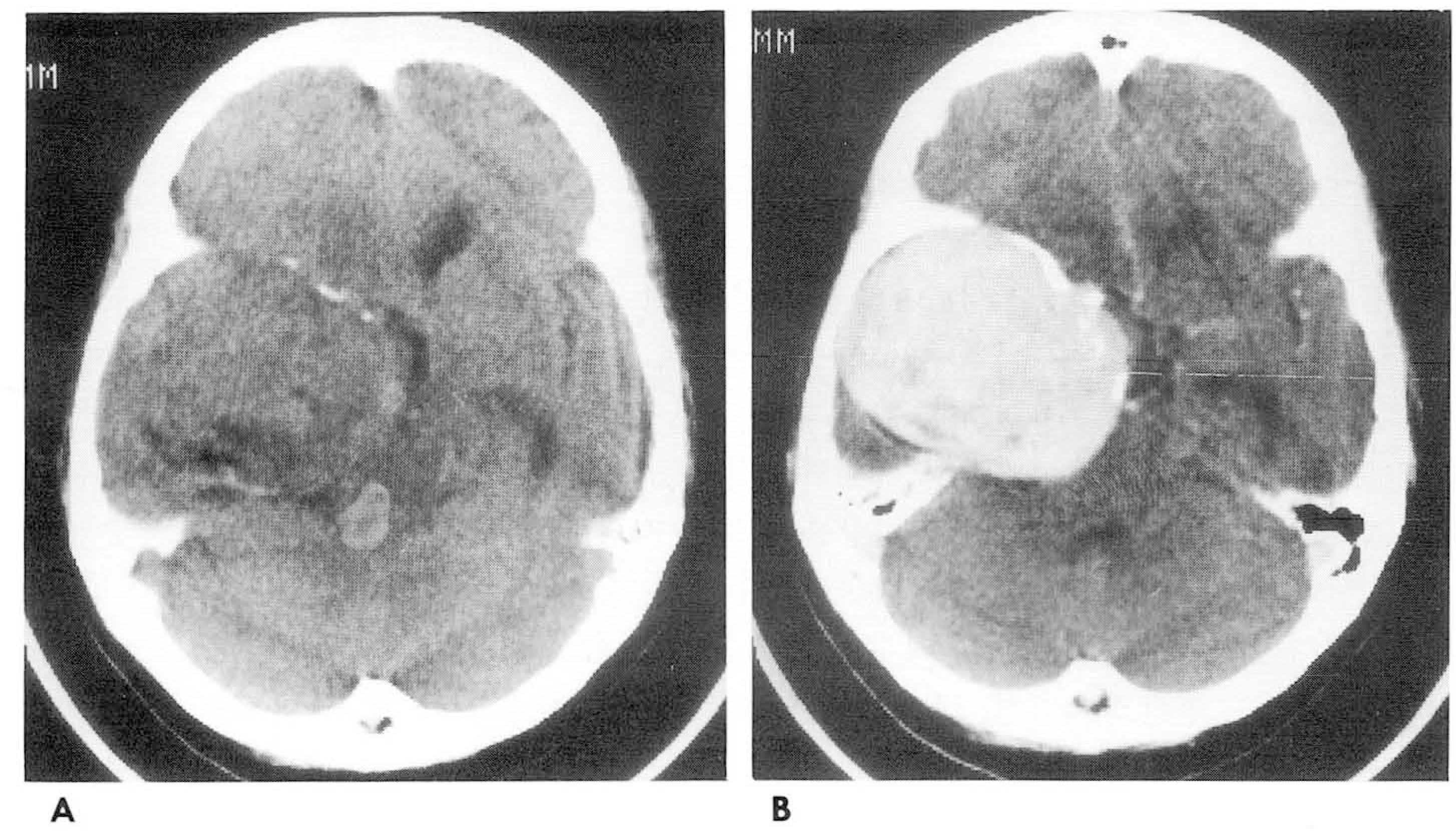
Fig. 1.
CT scan without (A) and with (B) intravenous contrast infusion. A. There is a relatively well demarcated heterogeneous low density mass in the right miaale cranial fossa. B. Intense and homogeneous enhancement is evident after contrast infusion.
jkrs-38-411f1.tif
Fig. 2.
MR imaging of the brain A. Tl-weighted image shows an extra-axial mass with mostly iso-intense signal and several hyperintense foci. B. T2-weighted image shows hyperintense signal. C. Gadolinium enhanced coronal Tl-weighted image shows homogeneous intense enhancement.
jkrs-38-411f2.tif
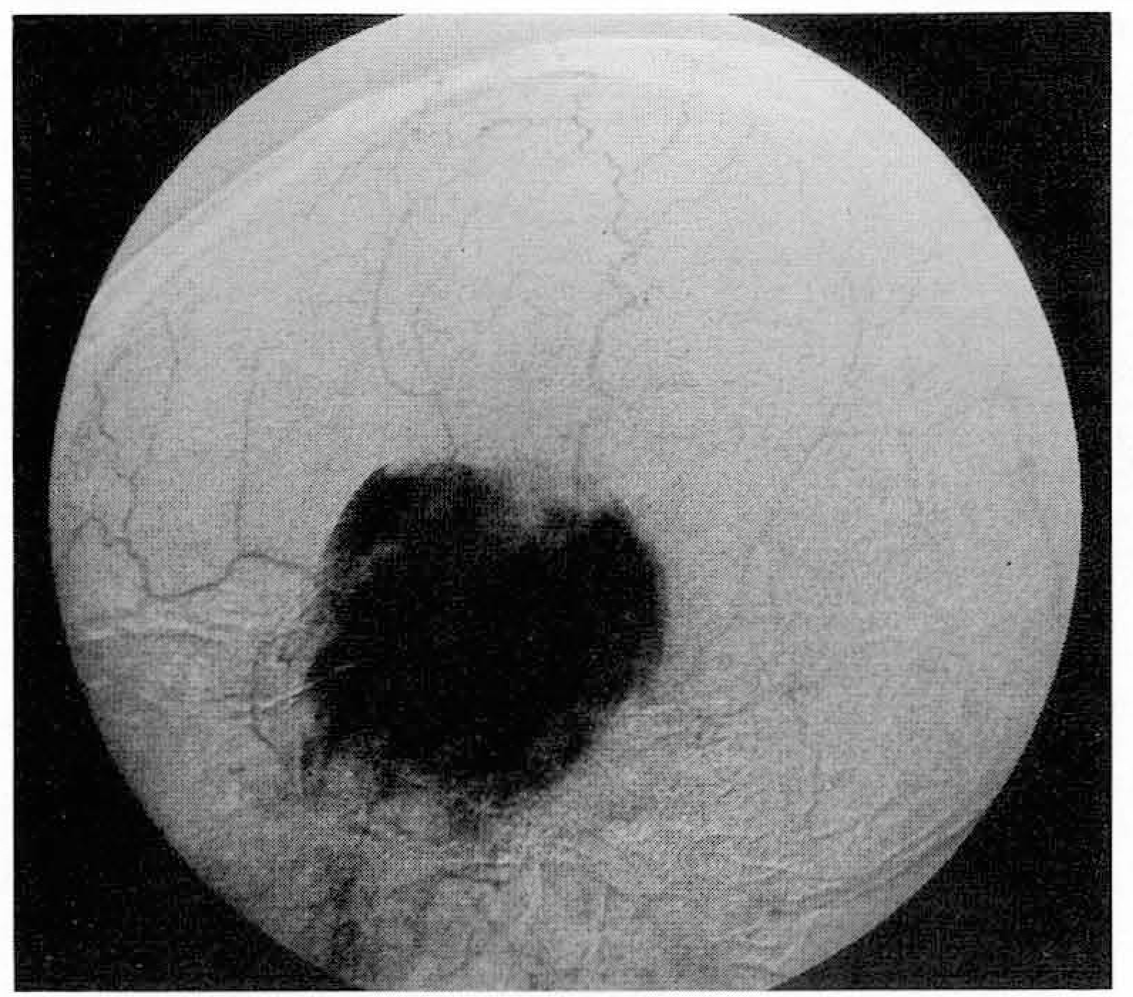
Fig. 3.
Selective right external carotid arteriogram shows dense capillary blush.
jkrs-38-411f3.tif
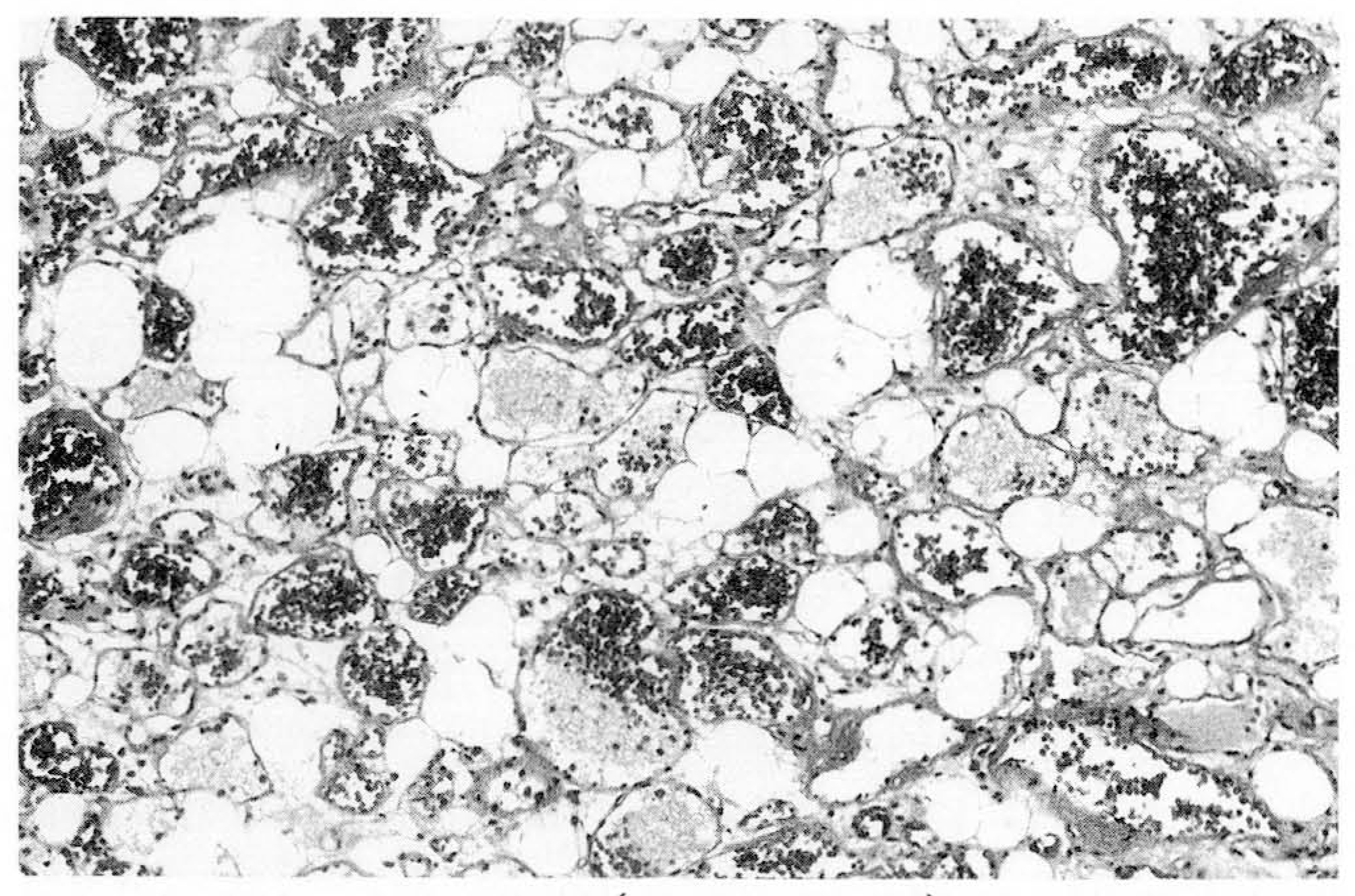
Fig. 4.
Photomicrograph(H&E, X 200) shows varying sized vessels and mature adipose tissue.
jkrs-38-411f4.tif
1998亼䰼 䴀⪤䏗㽔㻠㢨⪧⥴ 㛯㥬⩷㽔 㢄㟫 㪸佌 (Π)
䴀⥴ 䕻 㢄 㟫 㟤㉤㠀/古㜼㡛㹴
佌 㥗 䘸台㢄 / 㢄㟫
‘99 㡛⪧㼣㻠䏤 㴠㍓ ‘99 Fellowship 㴠㍓ 䘸台 98. 7. 31(凸) ⪧⥴ 劓㟤⧯䟛㣼㤰⥴
‘99 Scholarship 㴠㍓ 䘸台 98. 7. 31 (凸) ⪧⥴ 劓㟤⧯䟛㣼㤰⥴
㟤 54㡰 ⪧㷠䴀⥴ 䏱 ㋣⥴ ㋸䞣㟤㉤䘸台 98. 7. 31 (凸) ⪧⥴ 㽔䓌劓
㟼㴤㨻㍌ Booth 㴠㍓ 䘸台 98. 7. 31 (䕗) ⪧⥴ 㽔䓌劓
㽔㟼䬏䞣 䘸台 98. 8. 31(㤬) ⪧⥴ 㽔䓌劓
㟤 54㏘ ⪧㷠䴀⥴ 䏱 ㋣⥴ 98. 10. 15(䕗)-10. 17(⹠) ⧈⺬䞑䲐㤬䬤
5th AFSUMB 5th AFSUMB ⪧⥴ 98. 10. 23(凸)-27(⦬) Taipei, TAIWAN
㜄匼㞐㢴⳴⪧㷠䴀⥴ ㋸䞣㟤㉤䘸台 98. 10. 10(⹠) ㋸㢴⳴⪧⥴ 㽔䓌劓
’98亼䰼 ㉬匼㋸㢴⳴⪧㷠䴀⥴ 98. 11. 14(⹠) 䐈㟫
㟤 42 㡰㟼䓈㢨㴤⨨ 㢯㴤㤰㻤 㟯㷨 98.11. 6(凸)-13(凸) 㧸㟫
㟤 41 ㏘ 㟼䓈㢨匠㴤 99. 1. 䐈㟫
84th RSNA ㎼⼣䯤㋸䞣䕗䞣㟤㉤䘸台 98. 9. 30(⦬) ⪧⥴ 㽔䓌劓
84th RSNA(䉿䐈䏗㽔㻠㢨 ⪧⥴) 98. 11. 29(㢄)-12. 4(凸) Chicago. USA
㦈劓⪧⥴㙀 䏤⮤亄䓈 㦈劓⪧⢔㙀 䏤⮤亄䓈 䕗䞣 㟤㉤ 䘸台 1ô4’ 7, 10㤬䘰冴㙀(3古㤬 ㆨ) ⪧⥴ 㽔䓌劓
Imaging Conference ⢷䊀 䏗㽔㻠匄⪧ 98. 1. 21 (㷨) 18:00-20:00 㽄㻏㻤㥈䌯㤰(㧸㟫)
㹴⦬凐 䏗㽔㻠匄⪧ 98. 2. 18(㷨)
㴠千 䏗㽔㻠匄⪧ 98. 3. 18(㷨)
刄ô匘南 䏗㽔㻠匄⪧ 98. 5. 9(⹠) “
㛯㡔㟿 䏗㽔㻠匄⪧ 98. 5. 20(㷨) ”
㹴㪼 䏗㽔㻠匄⪧ 98. 6. 17(㷨)
䆼丘㼣㴣凐 䏗㽔㻠匄⪧ 98. 7. 8(㷨) “
㣠䏗 䏗㽔㻠匄⪧ 98. 9. 16(㷨)
⢷䊀 䏗㽔㻠匄⪧ 98. 10. 21(㷨)
㹴⦬凐 䏗㽔㻠匄⪧ 98. 11. 18(㷨) ”
㴠千 䏗㽔㻠匄⪧ 98. 12. 16(㷨〉
⢷䊀䏗㽔㻠匄⪧ 㨐劔⥴ subspecialty Meeting Case Discussion 98. 1. 14(㷨) 㻤㥈㛯㪧䌯㤰
2〜8㤬 䏱 11 㤬 㟫凐䕘㡼 䘜㤬 㺵㘈 䕗㥬㢄 㻤㥈䴀⪧劰䌯㤰
㙀䏗䕘㡼 98. 9. 19(⹠) 㟼佘䴀⪧劰䌯㤰?
Subspecialty meeting Interesting and Informative Cases for Residents 98. 10. 14(㷨) 㻤㥈㛯㪧䌯㤰?
㹟亼 Symposium 98. 12. 19(⹠) 㻤㥈㛯㪧䌯㤰?
䌋䊀䏗㽔㻠匄⪧ 㨐劔⥴ 㟫凐 㤬䟀䕘㡼 98. 1. 8(䕗)ô3. 12(䕗) 4. 9(䕗)ô5. 14(䕗) 7. 9(䕗)ô8. 13(䕗) 9. 10(䕗)ô10. 8(䕗) 1~ 5㤬 㻤㥈䴀 7~10㤬 㻤㥈㛯㪧
Subspecialty Conference 98. 2. 18(㷨) 㽄㻏㢨䜴㤰
㉨匼 Symposium 98. 5. 30(⹠) 㟼䉿㢨䴀
Subspecialty Conference 98. 11. 18(㷨) 㽄㻏㢨䜴㤰
㹟亼 Symposium 98. 12. 12(⹠) 㻤㥈䴀⪧䌯㤰
TOOLS
Similar articles