

 PDF
PDF ePub
ePub Citation
 Print
Print


Abstract
Purpose:
To evaluate the utility of fluid-attenuated inversion recovery (FLAIR) sequence by comparing the signal intensities in various cerebral lesions with those on T2-weighted MR imaging.
Materials & Methods:
In 41 patients who showed different signal intensities between Τ2-weighted images and FLAIR sequences, we reviewed the Virchow-Robin space(VRS), acute or chronic infarctions including lacunar cavities, and postoperative encephalomalacia. In all patients, the location, shape and size of abnormal signal intensities were evaluated.
Results:
The hyperintensities of VRS and lacunar infarctions on T2-weighted imaging appeared as hypointensities on FLAIR imaging. The hyperintense rims or crescents around lacunar cavities were only detected on FLAIR imaging. The extent of acute and chronic infarctions with homogenous hyperintensities seen on Τ 2-weigh ted images was well delineated on FLAIR imaping. Postoperative encephalomalacia and adjacent lesions showed low and high signal intensities, respectively, on FLAIR imaging, though they were hyperintense on T2-weighted images.
Go to : 
REFERENCES
1.이상현, 장기현, 박홍석등. 뇌질환의진단에있어서FLAIRMR sequence의임상적유용성. 대한방사선의학희지. 1997. 37:1–7.
2.Zawadzki MB., Atkinson D., Debtrick Μ, et al. Fluid-Attenuated Inversion Recovery (FLAIR) for assessment of cerebral infarction. Stroke. 1996. 27:1187–1191.

3.Coene BD., Hanjnal JV., Pennock JM, et al. MRI of the brain stem using fluid-attenuated inversion recovery pulse sequences. Neuroradiology. 1996. 35:327–331.
4.Alexander JA., Sheppard S., Davis PC, et al. Adult Cerebrovascular Diease: Role of Modified Rapid Fluid-Attenuated Inversion-Recovery Sequence. AJNR. 1996. 17:1507–1513.
5.Tsuchiya K., Mizutani Y., Hachiya J. Preliminary Evaluation of Fluid-Attenuated Inversion-Recovery MR in the Diagnosis of intracranial tumors. AJNR. 1996. 17:1081–1086.
6.Rydberg JN., Hammond CA., Grim RC, et al. Initial Clinical Experience in MR Imaging of the Brain with a Fast Fluid-attenuated Inversion-Recovery Pulse Sequence. Radiology. 1994. 193:173–180.
7.Hanjnal JV., Brant DJ., Kasuboskier L, et al. Use of fluid-attenuated inversion recovery (FLAIR) pulse sequence in MRI of the brain. J Comput Assist Tomogr. 1992. 16(6):841–844.
8.Coene BD., Hajnal JV., Gatehouse Peter, et al. MR of the brain using Fluid-Attenuated Inversion-Recovery (FLAIR) pulse sequence. AJNR. 1992. 13:1555–1564.
9.Simonson TM., Magnotta VA., Ebrabradt JC, et al. Echo-planar FLAIR imaging in evaluation of intracranial lesions. Radio Graphics. 1996. 16:575–584.
10.공근영, 최우석, 김의종. 뇌경색의급속FLAIR MR 영상소견: T2 강조영상과의비교. 대한방사선의학회지. 1997. 37:9–15.
11.Ogawa Τ., Okudera Τ., Fukasawa Ηet al. Unusual widening of Virchow-Robin space: MR appearance: AJNR. 1995. 6(16):1238–1242.
12.Wallace CJ., Sevick RJ. Multifocal white matter lesions. Semin Ultrasound CT MR. 1996. 17(3):251–264.
13.Linda A. Heier., Cristel J., Bauer ., Larry Schwartz, et al. Large Virchow-Robin spaces: MR-clinical correlation. AJNR. 1989. 10:929–936.
14.Braffman BH., Zimmerman RA., Trojanwski JQ, et al. Brain MR: Pathologic correlation with gross and histopathology 1. Lacunar infarction and Virchow-Robin spaces. AJNR. 1988. 9:621–628.
Go to :
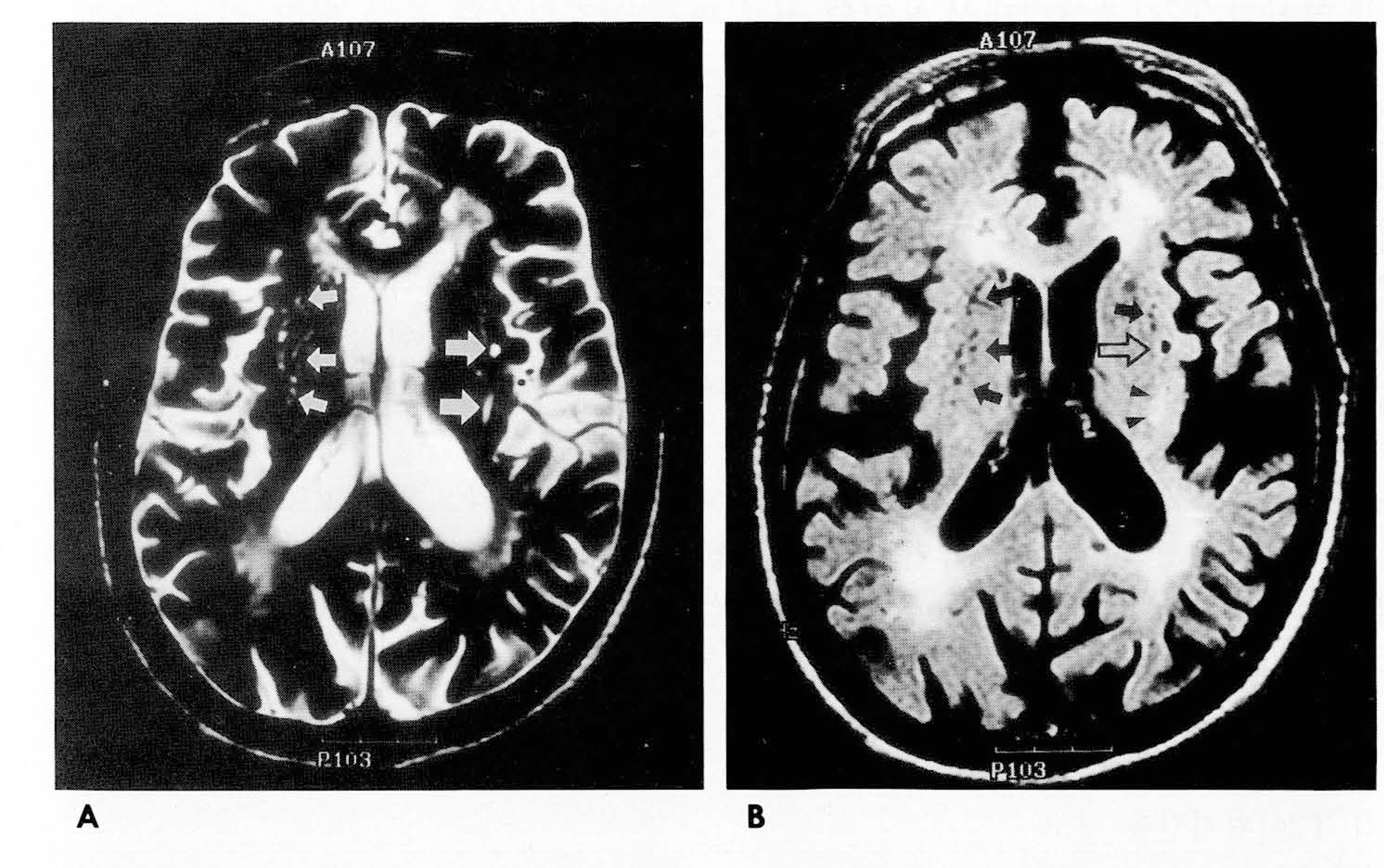 | Fig. 1.Dilated perivascualr space and ischemic lesions in 76-years-old man. A. Axial Τ 2-weighted MR image shows multiple variable-shaped hy- perintense lesions in both basal ganglia(small & large arrows). B. FLAIR image shows tubular or ovoid hypointense perivascualr spaces (solid arrows) and round hypointense lacunar infarction with peripheral hyperintensity(open arrow). The linear hyperintense ischemic lesion at the posterior aspect of left basal ganglia, which is seen in jig. 1A appears hyperintense(arrowhead- s) on FLAIR image. |
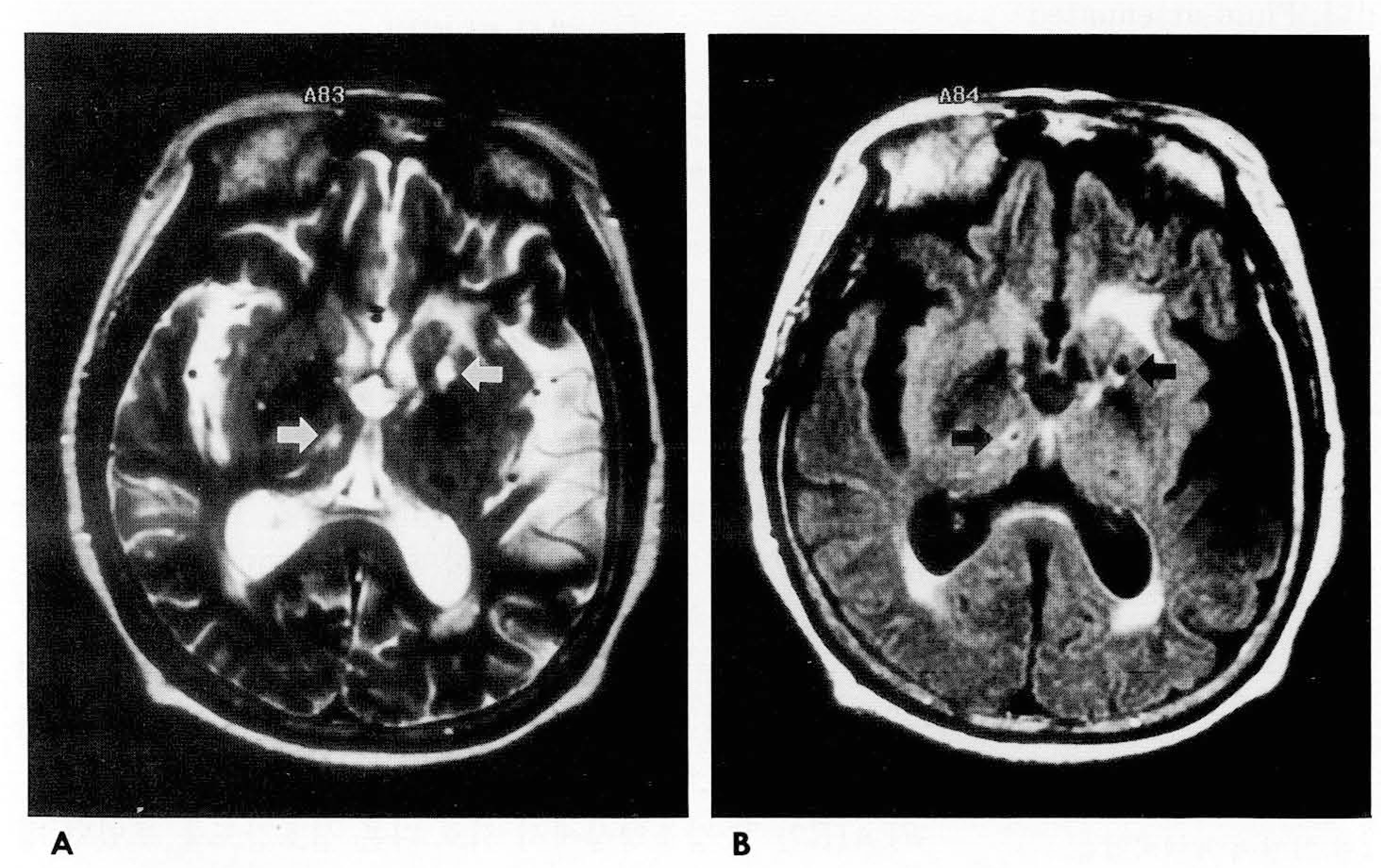 | Fig. 2.Lacunar infarction in 80- years-old woman. A. Axial Τ2-weighted image shows two round hyperintensities in left basal ganglia and right thalamus (arrows). B. FLAIR image shows small low signal intensities with hyperintense rim or crescent(arrows). |
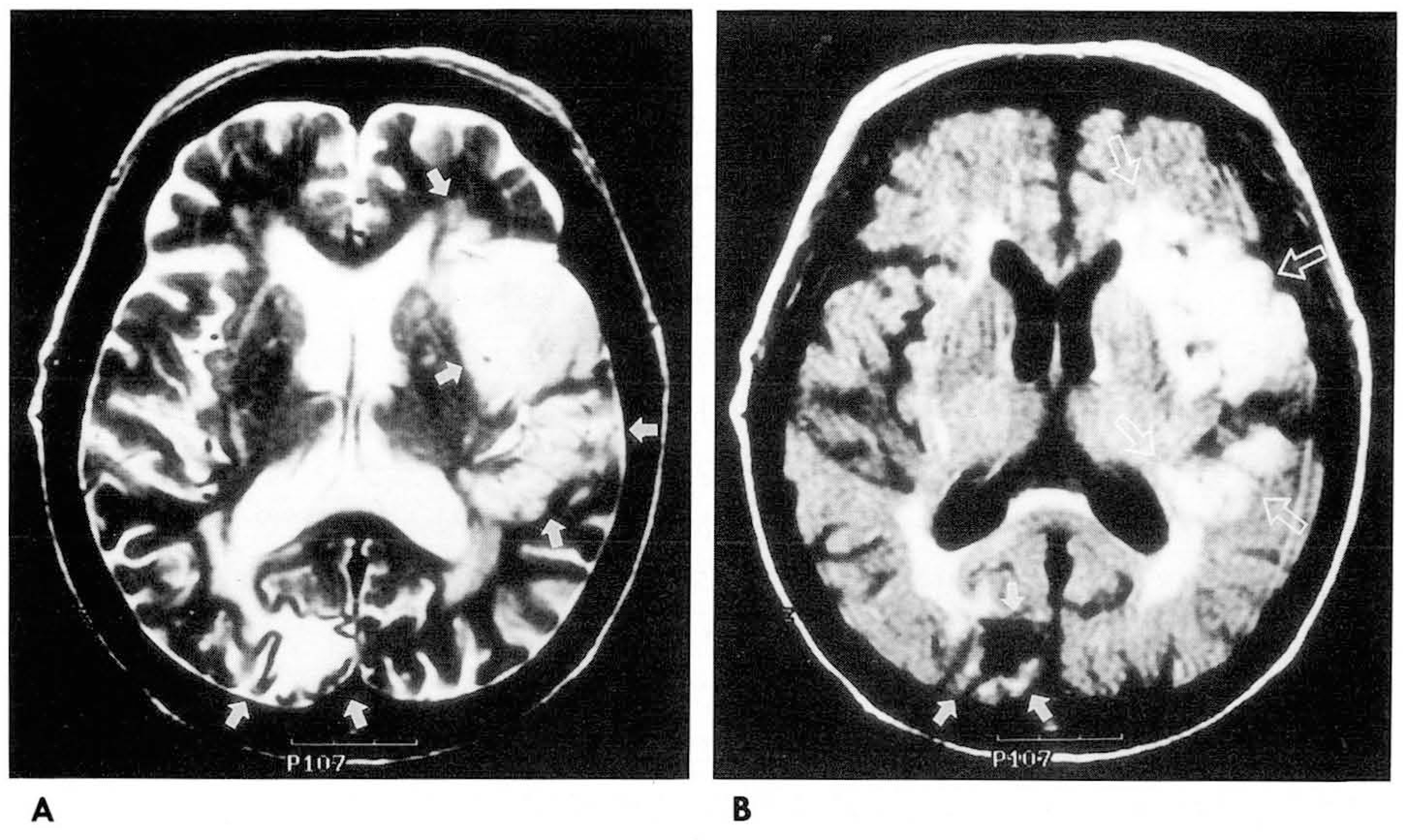 | Fig. 3.A 88-years-old woman with acute and chronic cerebral infarctions. A. Axial T2-weighted image shows cerebral infarctions in left frontal and parietal lobes, and right occipital lobe(arrows). B. On FLAIR image, the extent of acute(open arrows) and chronic (solid arrows) infarctions is well delineated. |
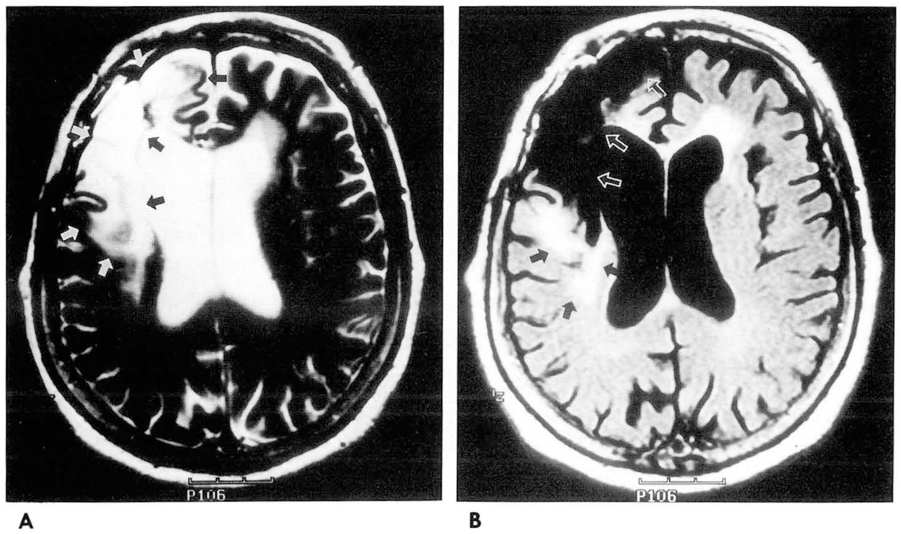 | Fig. 4.A 80-years-old woman with postoperative state of hematoma. A. Axial T2-weighted image shows huge hyperintense lesion in right frontal and temporal lobes(black and white arrows). B. On FLAIR image, brain tissue loss(white open arrows)and peripheral hyperintensity (black arrows) in the posterior aspect of the main lesion are seen. |
1998亼䰼
䴀⪤䏗㽔㻠㢨⪧⥴ 㛯㥬⩷㽔 㢄㟫 㪸佌 (I)
 XML Download