Abstract
Juvenile temporal arteritis (JTA) is a localized nodular arteritis confined to the temporal artery without evidence of systemic inflammation, and it occurs mainly in patients younger than 50 years. From the first case report, the pathological features of JTA have been suspected to be the morphological equivalent of Kimura disease (KD), which has been supported further by the concurrent cases of JTA with KD. We present the first case of bilateral JTA accompanying KD, which was confirmed by histological and ultrasound evaluations and supports the hypothesis that JTA is a manifestation of KD. The un-excised JTA lesion was resolved completely after corticosteroid therapy with no recurrence.
REFERENCES
1. Nesher G, Oren S, Lijovetzky G, Nesher R. Vasculitis of the temporal arteries in the young. Semin Arthritis Rheum. 2009; 39:96–107.

2. Watanabe C, Koga M, Honda Y, Oh-I T. Juvenile temporal arteritis is a manifestation of Kimura disease. Am J Dermatopathol. 2002; 24:43–9.

3. Brown I, Adkins G, McClymont K. Juvenile temporal arteritis: a case report. Pathology. 2005; 37:559–60.

4. Fukunaga M. Juvenile temporal arteritis associated with Kimura's disease. APMIS. 2005; 113:379–84.
5. Kim MB, Shin DH, Seo SH. Juvenile temporal arteritis with perifollicular lymphoid proliferation resembling Kimura disease. Report of a case. Int J Dermatol. 2011; 50:70–3.

6. Sun QF, Xu DZ, Pan SH, Ding JG, Xue ZQ, Miao CS, et al. Kimura disease: review of the literature. Intern Med J. 2008; 38:668–72.

7. Kolman OK, Spinelli HM, Magro CM. Juvenile temporal arteritis. J Am Acad Dermatol. 2010; 62:308–14.

8. Durant C, Connault J, Graveleau J, Toquet C, Brisseau JM, Hamidou M. Juvenile temporal vasculitis: a rare case in a middle-aged woman. Ann Vasc Surg. 2011; 25:384.e5-7.

9. McGeoch L, Silecky WB, Maher J, Carette S, Pagnoux C. Temporal arteritis in the young. Joint Bone Spine. 2013; 80:324–7.

10. Bollinger A, Leu HJ, Brunner U. Juvenile arteritis of ex-tracranial arteries with hypereosinophilia. Klin Wochenschr. 1986; 64:526–9.

11. Lie JT. Bilateral juvenile temporal arteritis. J Rheumatol. 1995; 22:774–6.
12. Fujimoto M, Sato S, Hayashi N, Wakugawa M, Tsuchida T, Tamaki K. Juvenile temporal arteritis with eosinophilia: a distinct clinicopathological entity. Dermatology. 1996; 192:32–5.

13. Arnander MW, Anderson NG, Schönauer F. The ultrasound halo sign in angiolymphoid hyperplasia of the temporal artery. Br J Radiol. 2006; 79:e184–6.

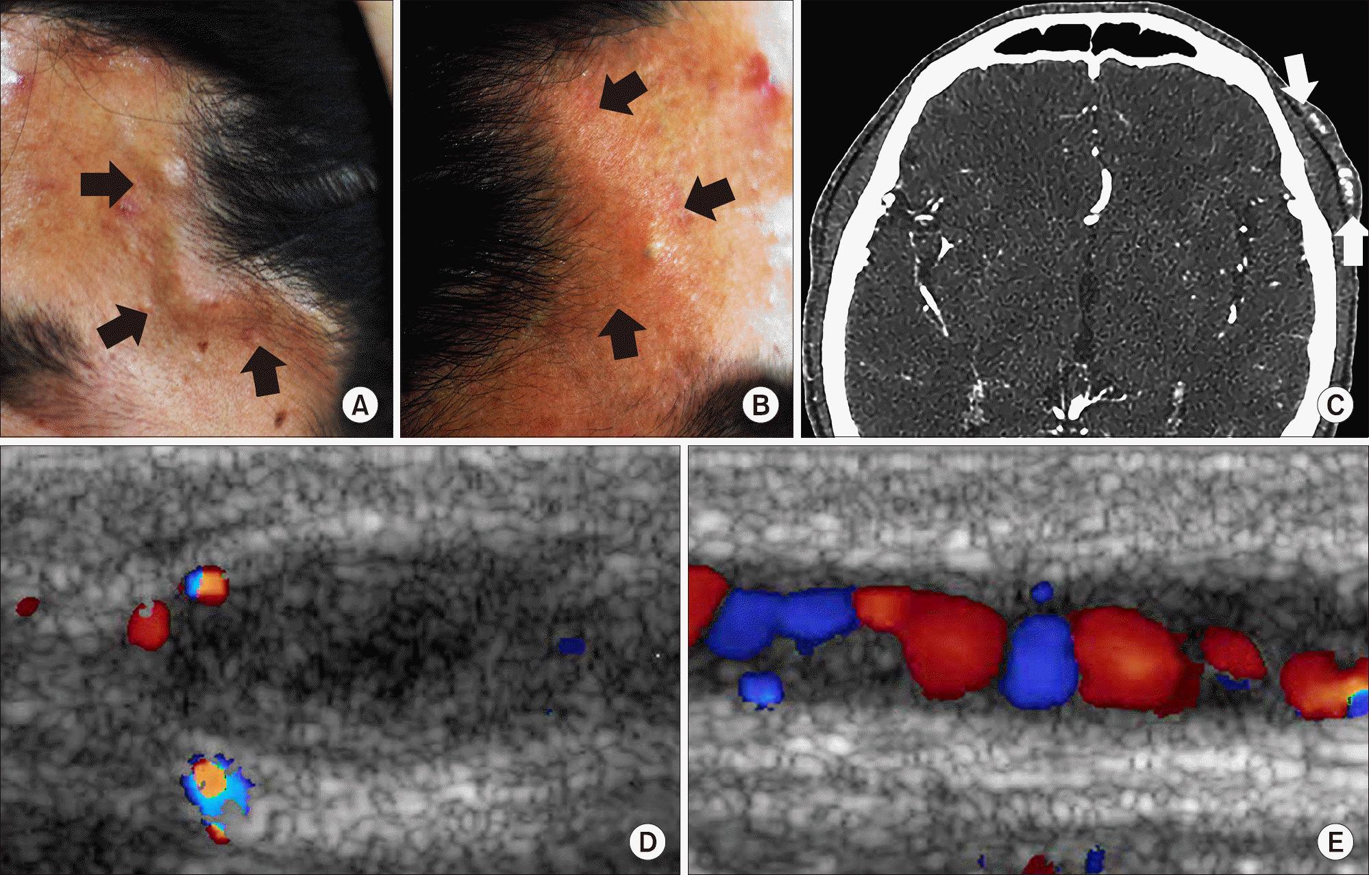
Figure 1.
Gross appearance (A: left, B: right), computed tomographic angiography (C) and Doppler ultrasound images (D: short axis, E: long axis) of the nodular lesion on the left superficial temporal artery.


 PDF
PDF ePub
ePub Citation
 Print
Print


 XML Download