

 PDF
PDF ePub
ePub Citation
 Print
Print


Abstract
Polyarteritis nodosa (PAN) has a broad spectrum of clinical presentation, since it affects small and medium-sized muscular arteries with microaneurysm formation, aneurysmal rupture with hemorrhage, thrombosis, and, consequently, organ ischemia or infarction. Although skeletal muscle involvement is well documented in patients with PAN, it can mimic more common diseases, and cause confusion and delays in diagnosis. PAN muscular involvement may have limited or early systemic forms with a benign course and excellent clinical response to corticosteroid therapy. Herein, we describe the clinical course and outcome of four unusual cases of PAN manifested by acute onset of pain and pitting edema in both lower extremities; in addition, we reviewed the relevant literature.
REFERENCES
1. Mahr A, Guillevin L, Poissonnet M, Aymé S. Prevalences of polyarteritis nodosa, microscopic polyangiitis, Wegener's granulomatosis, and Churg-Strauss syndrome in a French urban multiethnic population in 2000: a capture-recapture estimate. Arthritis Rheum. 2004; 51:92–9.

2. Guillevin L, Mahr A, Callard P, Godmer P, Pagnoux C, Leray E, et al. French Vasculitis Study Group. Hepatitis B virus-associated polyarteritis nodosa: clinical characteristics, outcome, and impact of treatment in 115 patients. Medicine (Baltimore). 2005; 84:313–22.
3. Bae YD, Choi HJ, Lee JC, Park JJ, Lee YJ, Lee EB, et al. Clinical features of polyarteritis nodosa in Korea. J Korean Med Sci. 2006; 21:591–5.
4. Pagnoux C, Seror R, Henegar C, Mahr A, Cohen P, Le Guern V, et al. Clinical features and outcomes in 348 patients with polyarteritis nodosa: a systematic retrospective study of patients diagnosed between 1963 and 2005 and entered into the French Vasculitis Study Group Database. Arthritis Rheum. 2010; 62:616–26.
5. Plumley SG, Rubio R, Alasfar S, Jasin HE. Polyarteritis nodosa presenting as polymyositis. Semin Arthritis Rheum. 2002; 31:377–83.
6. Kamimura T, Hatakeyama M, Torigoe K, Nara H, Kaneko N, Satou H, et al. Muscular polyarteritis nodosa as a cause of fever of undetermined origin: a case report and review of the literature. Rheumatol Int. 2005; 25:394–7.
7. Khellaf M, Hamidou M, Pagnoux C, Michel M, Brisseau JM, Chevallier X, et al. Vasculitis restricted to the lower limbs: a clinical and histopathological study. Ann Rheum Dis. 2007; 66:554–6.
8. Gallien S, Mahr A, Réty F, Kambouchner M, Lhote F, Cohen P, et al. Magnetic resonance imaging of skeletal muscle involvement in limb restricted vasculitis. Ann Rheum Dis. 2002; 61:1107–9.
9. Kim YJ, Kim SI, Hong KW, Kang MW. Diagnostic value of 18F-FDG PET/CT in patients with fever of unknown origin. Intern Med J. 2012; 42:834–7.
10. Lee LM, Moloney PJ, Wong HC, Magil AB, McLoughlin MG. Testicular pain: an unusual presentation of polyarteritis nodosa. J Urol. 1983; 129:1243–4.
11. Guillevin L, Cohen P, Mahr A, Arène JP, Mouthon L, Puéchal X, et al. Treatment of polyarteritis nodosa and microscopic polyangiitis with poor prognosis factors: a prospective trial comparing glucocorticoids and six or twelve cyclophosphamide pulses in sixty-five patients. Arthritis Rheum. 2003; 49:93–100.
12. Ribi C, Cohen P, Pagnoux C, Mahr A, Arène JP, Puéchal X, et al. Treatment of polyarteritis nodosa and microscopic polyangiitis without poor-prognosis factors: A prospective randomized study of one hundred twenty-four patients. Arthritis Rheum. 2010; 62:1186–97.
Figure 1.
(A) Erythematous swelling in the right lower leg and (B) computed tomographic scan showed intramuscular central low density and peripheral enhancement, and superficial fas-cial thickening with subcutaneous fat edema (arrows) and (C) whole body positron emission tomography scan showed markedly increased fluorodeoxyglucose uptakes in bilateral lower leg muscles.
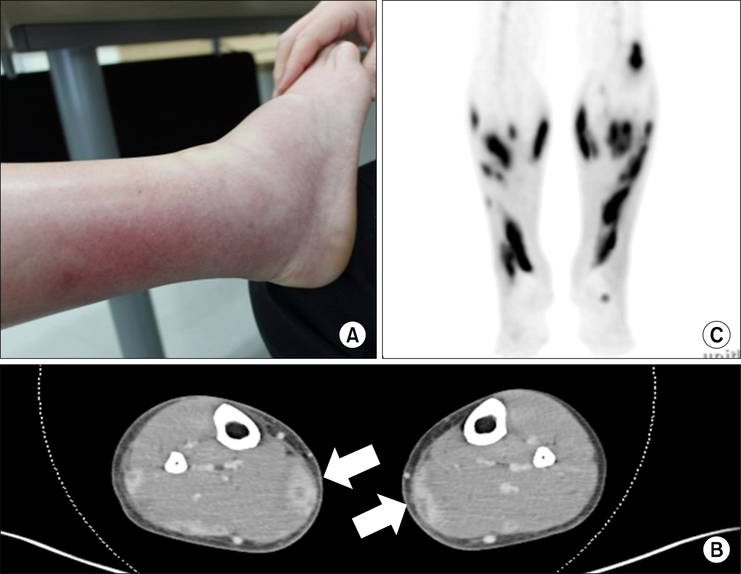
Figure 2.
(A) Axial T2-weighted magnetic resonance imaging revealed diffuse edematous change and enhancement in the right thigh muscles (arrows) and (B, C) infiltration of vessel walls by neutrophils were seen histologically (H&E stain).
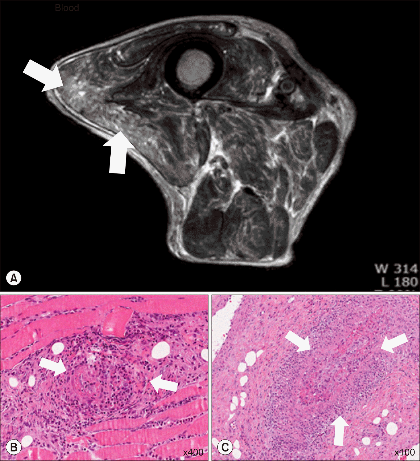
Figure 3.
(A) Erythematous macular rash on the left lower extremity and (B, C) axial T2-wei-ghted magnetic resonance imaging in the right lower leg showed multifocal ill-defined signal abnormality in the muscles and subcutaneous layers (arrows).
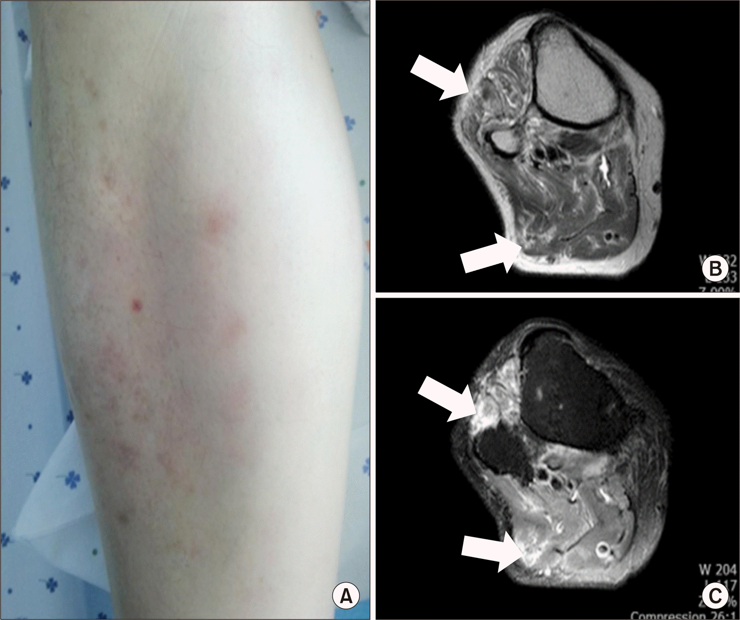
Figure 4.
Orbital computed tomographic scan revealed swelling and increased attenuation (arrow) of left periorbital soft tissue.
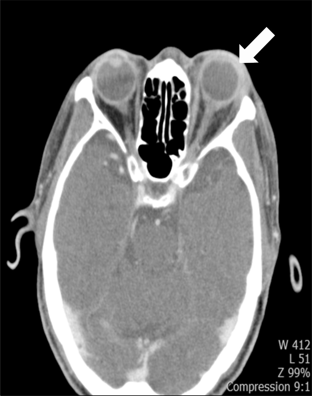
Table 1.
Summary of clinical presentations in our case series
 XML Download