Abstract
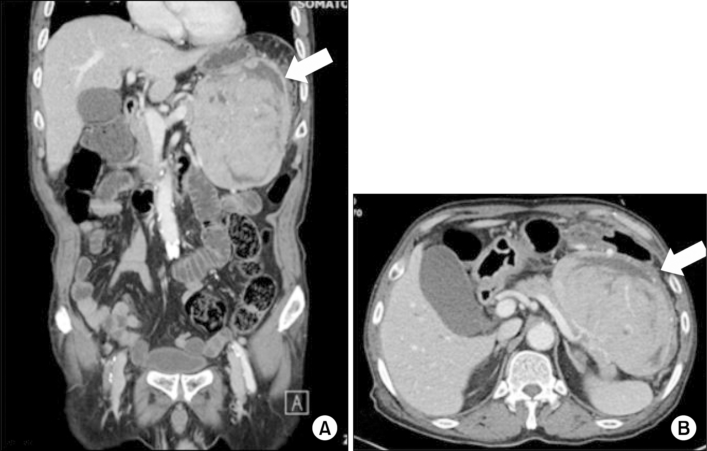
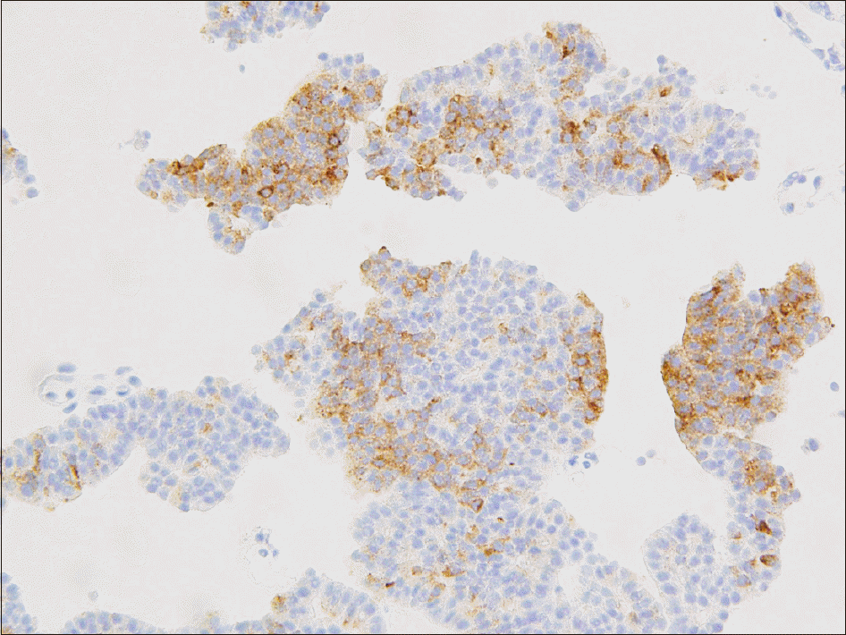
Pancreatic neoplasm is complicated and can be preceded by extra-pancreatic manifestations, such as cutaneous and musculoskeletal symptoms. Awareness of these associations is important for timely diagnosis and appropriate treatment. We report a case of pancreatic neuroendocrine tumor (NET) presenting with arthritis and panniculitis. The patient had a two month history of right knee pain and subcutaneous nodules in both legs. Synovial fluid analysis from the right knee joint revealed a mildly increased white blood cell count without crystallization. A skin biopsy of a subcutaneous nodule revealed lobular panniculitis. The initial treatment with empirical antibiotics did not alleviate the symptoms; however, the right knee arthritis and skin nodules improved with steroid treatment. On the eighth day of hospitalization, the patient complained of abdominal discomfort. Abdominopelvic computed tomography scanning revealed a 14-cm sized pancreatic mass with peritoneal metastasis. Percutaneous needle biopsy confirmed the diagnosis of pancreatic NET.
REFERENCES
1. Oh CM, Won YJ, Jung KW, Kong HJ, Cho H, Lee JK, et al. Cancer statistics in Korea: incidence, mortality, survival, and prevalence in 2013. Cancer Res Treat. 2016; 48:436–50.

2. Ruess DA, Makowiec F, Chikhladze S, Sick O, Riediger H, Hopt UT, et al. The prognostic influence of intrapancreatic tumor location on survival after resection of pancreatic ductal adenocarcinoma. BMC Surg. 2015; 15:123.

4. Shbeeb MI, Duffy J, Bjornsson J, Ashby AM, Matteson EL. Subcutaneous fat necrosis and polyarthritis associated with pancreatic disease. Arthritis Rheum. 1996; 39:1922–5.

5. Dahl PR, Su WP, Cullimore KC, Dicken CH. Pancreatic panniculitis. J Am Acad Dermatol. 1995; 33:413–7.

7. Lewis CT 3rd, Tschen JA, Klima M. Subcutaneous fat necrosis associated with pancreatic islet cell carcinoma. Am J Dermatopathol. 1991; 13:52–6.

8. Schwartz RA, Fleishman JS. Association of insulinoma with subcutaneous nodular fat necrosis. J Surg Oncol. 1981; 16:305–11.

9. Preiss JC, Faiss S, Loddenkemper C, Zeitz M, Duchmann R. Pancreatic panniculitis in an 88-year-old man with neuroendocrine carcinoma. Digestion. 2002; 66:193–6.

10. Berkovic D, Hallermann C. Carcinoma of the pancreas with neuroendocrine differentiation and nodular panniculitis. Onkologie. 2003; 26:473–6.

11. Szekanecz E, András C, Sándor Z, Antal-Szalmás P, Szántó J, Tamási L, et al. Malignancies and soluble tumor antigens in rheumatic diseases. Autoimmun Rev. 2006; 6:42–7.

12. Osborne RR. Functioning acinous cell carcinoma of the pancreas accompanied with widespread focal fat necrosis. Arch Intern Med (Chic). 1950; 85:933–43.

13. van Klaveren RJ, de Mulder PH, Boerbooms AM, van de Kaa CA, van Haelst UJ, Wagener DJ, et al. Pancreatic carcinoma with polyarthritis, fat necrosis, and high serum lipase and trypsin activity. Gut. 1990; 31:953–5.

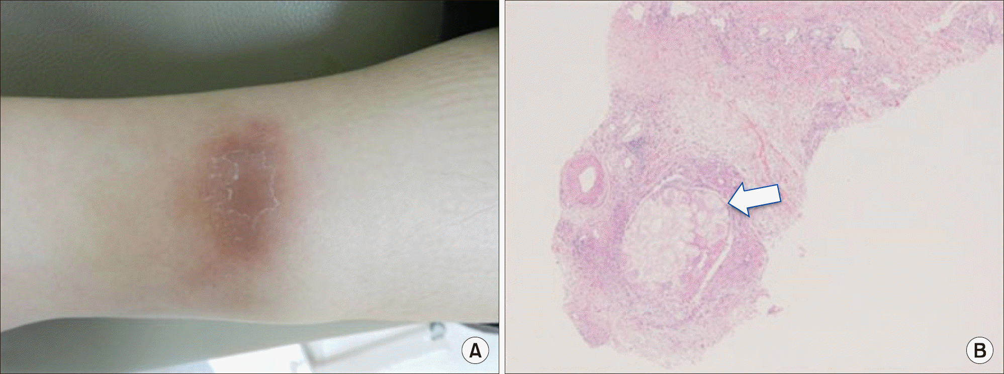
Figure 1.
(A) A subcutaneous lesion is present on the posterior surface of the right leg, indicating panniculitis. (B) Punch biopsy revealing lobular panniculitis with fat necrosis (H&E, x40, arrow).

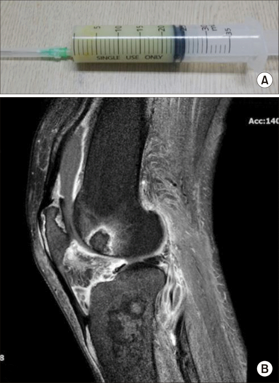
Figure 2.
(A) Aspirated right knee joint fluid showing turbid appearance. (B) Right knee magnetic resonance imaging showing fluid collection and multifocal enhancement around the knee joint.


 PDF
PDF ePub
ePub Citation
 Print
Print


 XML Download