Abstract
Objective
To analyze radiologic findings of cervical involvement in ankylosing spondylitis (AS) patients, determine its association with structural severity and clinical variables, and to divide radiologic findings of atlantoaxial ankylosis (AAA) in AS patients into three anatomical components.
Methods
The study includes 150 AS patients with either AAA (62 patients) or atlantoaxial subluxation (AAS, 88 patients) who underwent plain radiography of the cervical spine on flexion at our tertiary center for rheumatic diseases. The study subjects' medical records were reviewed. Lateral plain radiographs of the cervical spine were analyzed by a musculoskeletal radiologist. We compared the results of the modified Stoke Ankylosing Spondylitis Spinal Score (mSASSS) between AAS and AAA patients to determine if mSASSS was related to severity or duration of AS.
Results
The mean duration of illness in AS patients with AAA was 19.3 years, and in AAS patients 13.7 years (p<0.01). The mean total mSASSS of AS patients with AAA was 40.1, and of AAS patients 16.5 (p<0.001), and was positively associated with the development of AAA and AAS. The odds ratio (OR) of AAA development by cervical spine mSASSS change was higher (OR, 1.079) than the OR (1.049) of lumbar spine mSASSS even after adjusting for age, sex, and disease duration.
REFERENCES
1. Gouveia EB, Elmann D, Morales MS. Ankylosing spondylitis and uveitis: overview. Rev Bras Reumatol. 2012; 52:742–56.
2. Archer JR, Keat AC. Ankylosing spondylitis: time to focus on ankylosis. J Rheumatol. 1999; 26:761–4.
3. Martel W. The occipito-atlantoaxial joints in rheumatoid arthritis and ankylosing spondylitis. Am J Roentgenol Radium Ther Nucl Med. 1961; 86:223–40.
4. Hunter T. The spinal complications of ankylosing spondylitis. Semin Arthritis Rheum. 1989; 19:172–82.

5. Liang CL, Lu K, Lee TC, Lin YC, Chen HJ. Dissociation of atlantoaxial junction in ankylosing spondylitis: case report. J Trauma. 2002; 53:1173–5.

6. van der Linden S, Valkenburg HA, Cats A. Evaluation of diagnostic criteria for ankylosing spondylitis. A proposal for modification of the New York criteria. Arthritis Rheum. 1984; 27:361–8.
7. Kauppi M, Neva MH. Sensitivity of lateral view cervical spine radiographs taken in the neutral position in atlantoaxial subluxation in rheumatic diseases. Clin Rheumatol. 1998; 17:511–4.

8. Meijers KA, van Voss SF, François RJ. Radiological changes in the cervical spine in ankylosing spondylitis. Ann Rheum Dis. 1968; 27:333–8.

9. Lee HS, Kim TH, Yun HR, Park YW, Jung SS, Bae SC, et al. Radiologic changes of cervical spine in ankylosing spondylitis. Clin Rheumatol. 2001; 20:262–6.

10. El Maghraoui A, Bensabbah R, Bahiri R, Bezza A, Guedira N, Hajjaj-Hassouni N. Cervical spine involvement in ankylosing spondylitis. Clin Rheumatol. 2003; 22:94–8.
11. Lee JY, Kim JI, Park JY, Choe JY, Kim CG, Chung SH, et al. Cervical spine involvement in longstanding ankylosing spondylitis. Clin Exp Rheumatol. 2005; 23:331–8.
12. Sanzhang C, Rothschild BM. Zygapophyseal and costoverte-bral/costotransverse joints: an anatomic assessment of arthritis impact. Br J Rheumatol. 1993; 32:1066–71.

13. Simkin PA, Downey DJ, Kilcoyne RF. Apophyseal arthritis limits lumbar motion in patients with ankylosing spondylitis. Arthritis Rheum. 1988; 31:798–802.

14. Russell AS, Jackson F. Computer assisted tomography of the apophyseal changes in patients with ankylosing spondylitis. J Rheumatol. 1986; 13:581–5.
15. Laiho K, Kauppi M. The cervical spine in patients with ankylosing spondylitis. Clin Exp Rheumatol. 2002; 20:738.
Figure 1.
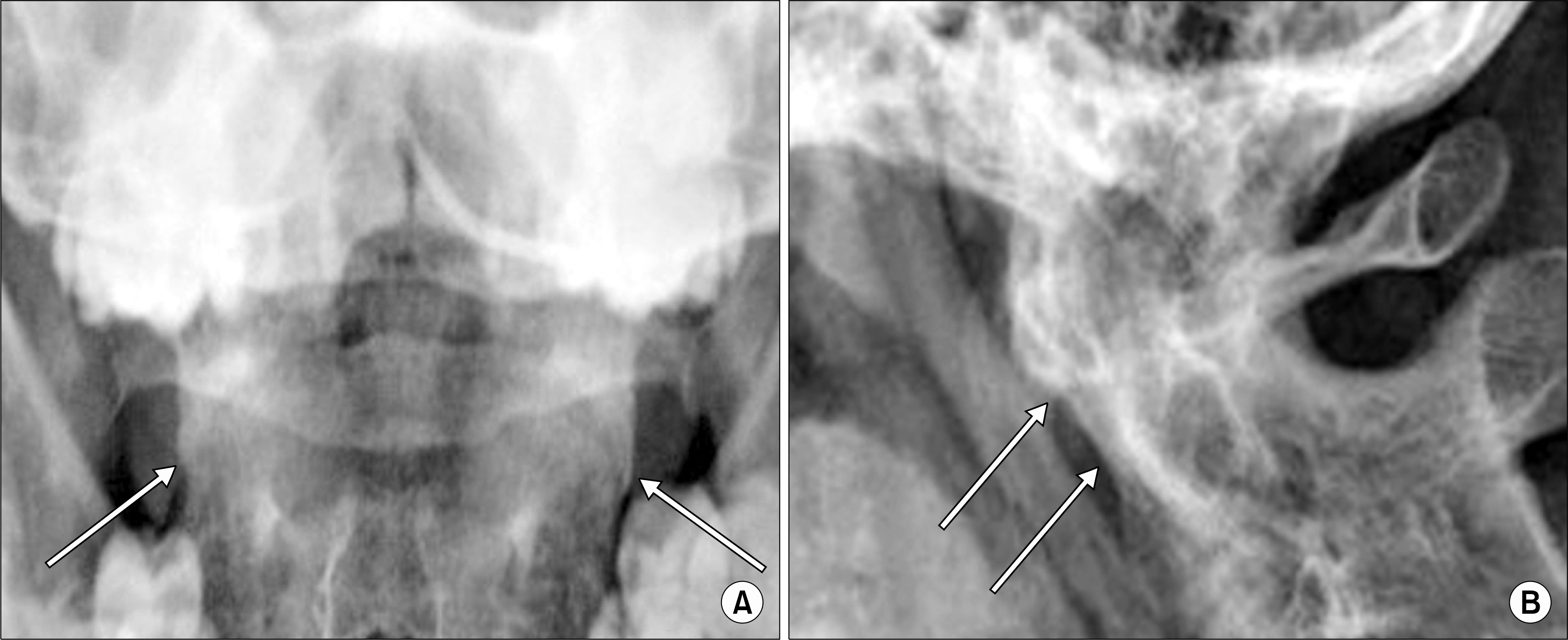
(A) A 31-year-old ankylosing spondylitis patient with duration of 9 years. An anteroposterior open-mouth view of the cervical spine shows obliteration of atlantoaxial facet joints (arrows, component II). (B) A lateral radiograph of the cervical spine with full flexion of the same patient shows bony ankylosis of the atlantoaxial joint space and ankylosis of anterior longitudinal ligament (arrows, component I and III).

Figure 2.
(A) A 34-year-old ankylosing spondylitis patient with a disease duration of 10 years. An anteroposterior open-mouth view of the cervical spine reveals obliteration of atlantoaxial facet joint (arrows, component II). (B) A lateral radiograph of the cervical spine with full flexion of the same patient shows sparing of the atlantoaxial joint space.

Figure 3.
(A) A 33-year-old ankylosing spondylitis patient with duration of 8 years. An anteroposterior open-mouth view of the cervical spine reveals sparing of the atlantoaxial facet joint. (B) A lateral radiograph of the cervical spine with full flexion of the same patient shows ankylosis of anterior longitudinal ligament (arrow, component III).

Table 1.
Basic demographic, clinical findings, and mSASSS of 150 patients
| Characteristic | Group A* (n=62) | Group B† (n=88) | p-value |
|---|---|---|---|
| Age (yr) | 40.1±8.2 | 34.6±10.9 | 0.001 |
| Sex, male | 61 (98.4) | 80 (90.9) | 0.081 |
| Disease duration (yr) | 19.3±7.7 | 13.7±9.0 | <0.001 |
| Ocular symptom | 19 (30.6) | 44 (50.0) | 0.206 |
| HLA-B27 positivity mSASSS | 61 (98.4) | 86 (97.7) | 1.000 |
| C-spine score | 22.6±12.4 | 10.1±9.6 | <0.001 |
| L-spine score | 17.5±18.9 | 6.4±11.2 | <0.001 |
| Total score | 40.1±25.3 | 16.5±18.1 | <0.001 |
Table 2.
Odds ratio of AAA development according to disease duration and the change of mSASSS (compared with development of AAS)
| Variable | Odds ratio | 95% confidence interval |
|---|---|---|
| Disease duration | ||
| Unadjusted | 1.081 | 1.037∼1.128 |
| Adjusted for age, sex | 1.065 | 1.010∼1.123 |
| Adjusted for age, sex and total mSASSS | 1.055 | 0.998∼1.116 |
| Change of mSASSS | ||
| C-spine∗ | 1.079 | 1.042∼1.118 |
| L-spine∗ | 1.049 | 1.018∼1.081 |


 PDF
PDF ePub
ePub Citation
 Print
Print


 XML Download