

 PDF
PDF ePub
ePub Citation
 Print
Print


Abstract
Takayasu's arteritis (TA), a granulomatous vasculitis, affects the aorta and its major branches. Glucocorticoids are an effective treatment for patients with active TA, but some patients fail to achieve or maintain remission with the conventional therapy, and side effects resulting from long-term glucocorticoid therapy are potentially serious. Anti-tumor necrosis factor-α agents, such as infliximab, may be efficient in patients with refractory TA. We report on a 24-year-old female patient with refractory TA who 18 F-fluoro-2-de-was treated successfully with infliximab. Clinical remission was induced as determined by repeated oxy-D-glucose positron emission tomography scans combined with assay of serological inflammatory markers.
Go to : 
REFERENCES
1. Kötter I, Henes JC, Wagner AD, Loock J, Gross WL. Does glucocorticosteroid-resistant large-vessel vasculitis (giant cell arteritis and Takayasu arteritis) exist and how can remission be achieved? A critical review of the literature. Clin Exp Rheumatol. 2012; 30(1 Suppl 70):S114–29.
2. Molloy ES, Langford CA, Clark TM, Gota CE, Hoffman GS. Anti-tumour necrosis factor therapy in patients with refractory Takayasu arteritis: long-term follow-up. Ann Rheum Dis. 2008; 67:1567–9.

3. Arend WP, Michel BA, Bloch DA, Hunder GG, Calabrese LH, Edworthy SM, et al. The American College of Rheumatology 1990 criteria for the classification of Takayasu arteritis. Arthritis Rheum. 1990; 33:1129–34.
4. Kerr GS, Hallahan CW, Giordano J, Leavitt RY, Fauci AS, Rottem M, et al. Takayasu arteritis. Ann Intern Med. 1994; 120:919–29.
5. Mekinian A, Néel A, Sibilia J, Cohen P, Connault J, Lambert M, et al. Club Rhumatismes et Inflammation, French Vasculitis Study Group and Société Nationale Française de Médecine Interne. Efficacy and tolerance of infliximab in refractory Takayasu arteritis: French multicentre study. Rheumatology (Oxford). 2012; 51:882–6.
6. Clifford A, Hoffman GS. Recent advances in the medical management of Takayasu arteritis: an update on use of biologic therapies. Curr Opin Rheumatol. 2014; 26:7–15.
7. Osman M, Pagnoux C, Dryden DM, Storie D, Yacyshyn E. The role of biological agents in the management of large vessel vasculitis (LVV): a systematic review and meta-analysis. PLoS One. 2014; 9:e115026.
8. Comarmond C, Plaisier E, Dahan K, Mirault T, Emmerich J, Amoura Z, et al. Anti TNF-α in refractory Takayasu's arteritis: cases series and review of the literature. Autoimmun Rev. 2012; 11:678–84.
9. Terao C, Yoshifuji H, Mimori T. Recent advances in Takayasu arteritis. Int J Rheum Dis. 2014; 17:238–47.
10. Tezuka D, Haraguchi G, Ishihara T, Ohigashi H, Inagaki H, Suzuki J, et al. Role of FDG PET-CT in Takayasu arteritis: sensitive detection of recurrences. JACC Cardiovasc Imaging. 2012; 5:422–9.
11. Lee KH, Cho A, Choi YJ, Lee SW, Ha YJ, Jung SJ, et al. The role of (18) F-fluorodeoxyglucose-positron emission tomography in the assessment of disease activity in patients with takayasu arteritis. Arthritis Rheum. 2012; 64:866–75.
Go to :
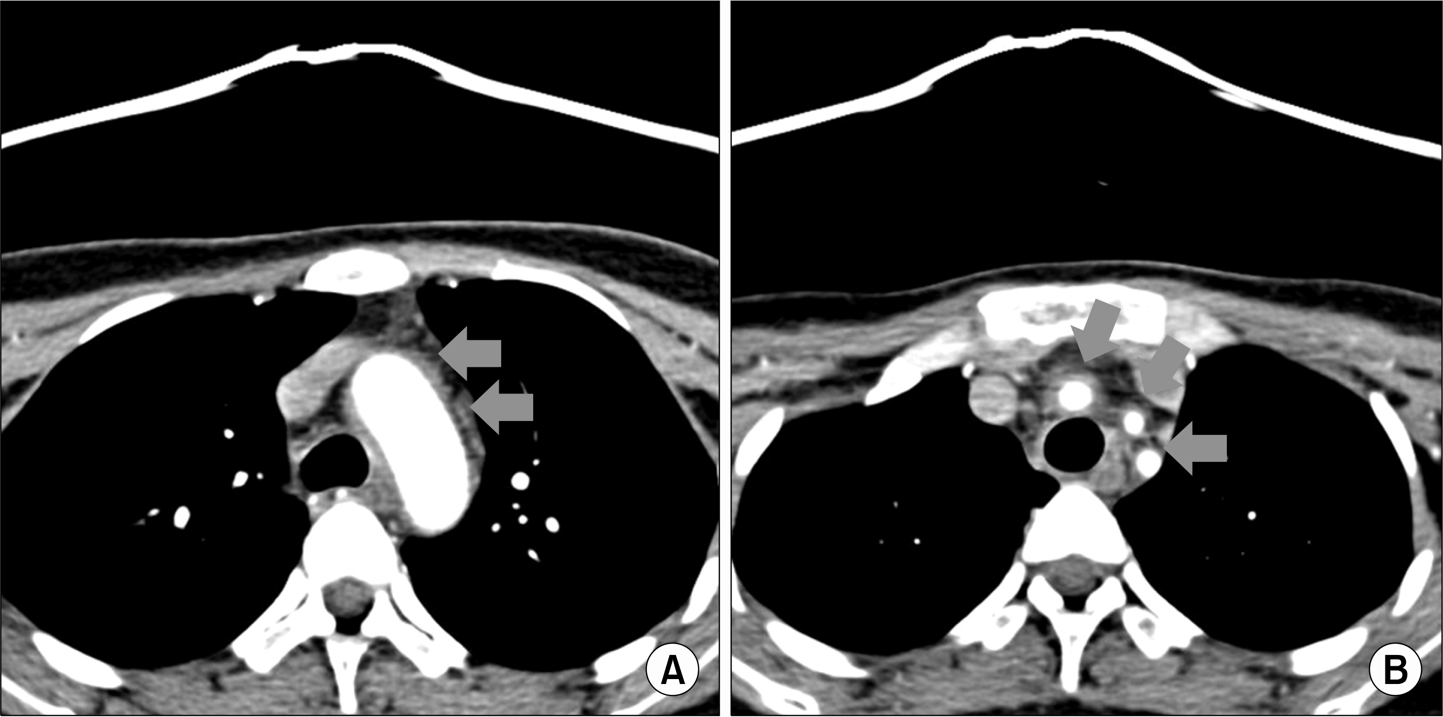 | Figure 1.Contrast-enhanced computed tomography scan shows diffuse wall thickening (arrows) in (A) aortic arch, and (B) the right brachiocephalic trunk, left common carotid and subclavian arteries. |
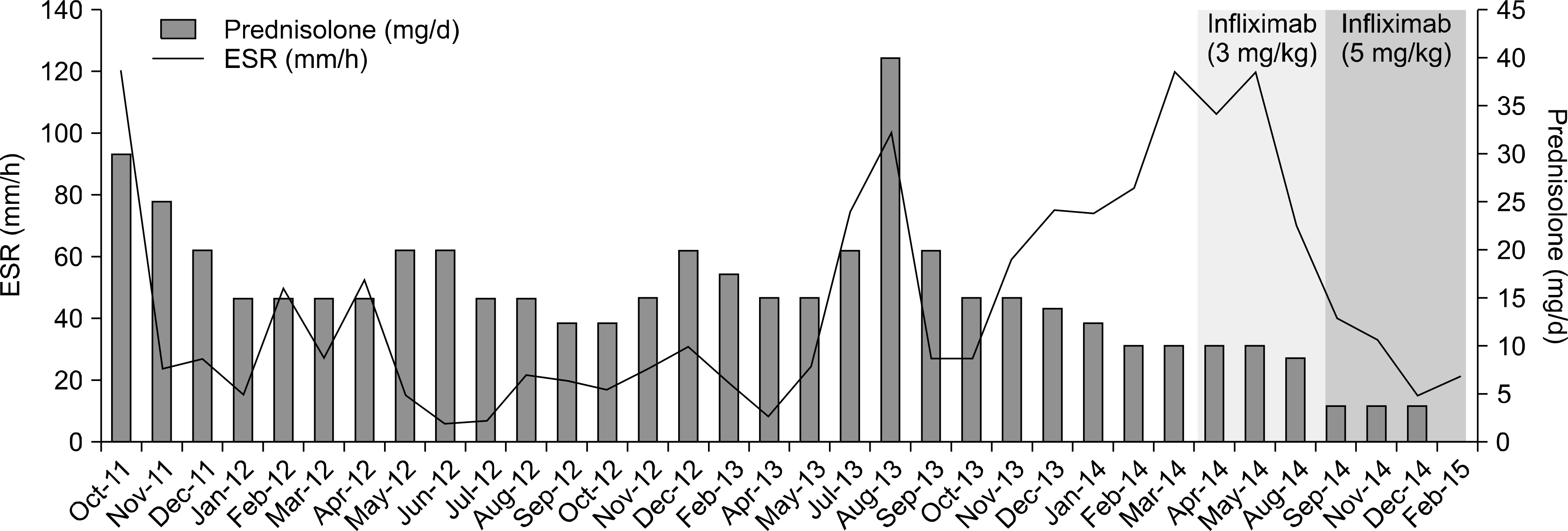 | Figure 2.Treatment timeline showing the response of systemic inflammatory markers to glucocorticoid and infliximab during the course of treatment. Erythrocyte sedimentation rate (ESR) denotes erythrocyte sediment rate. |
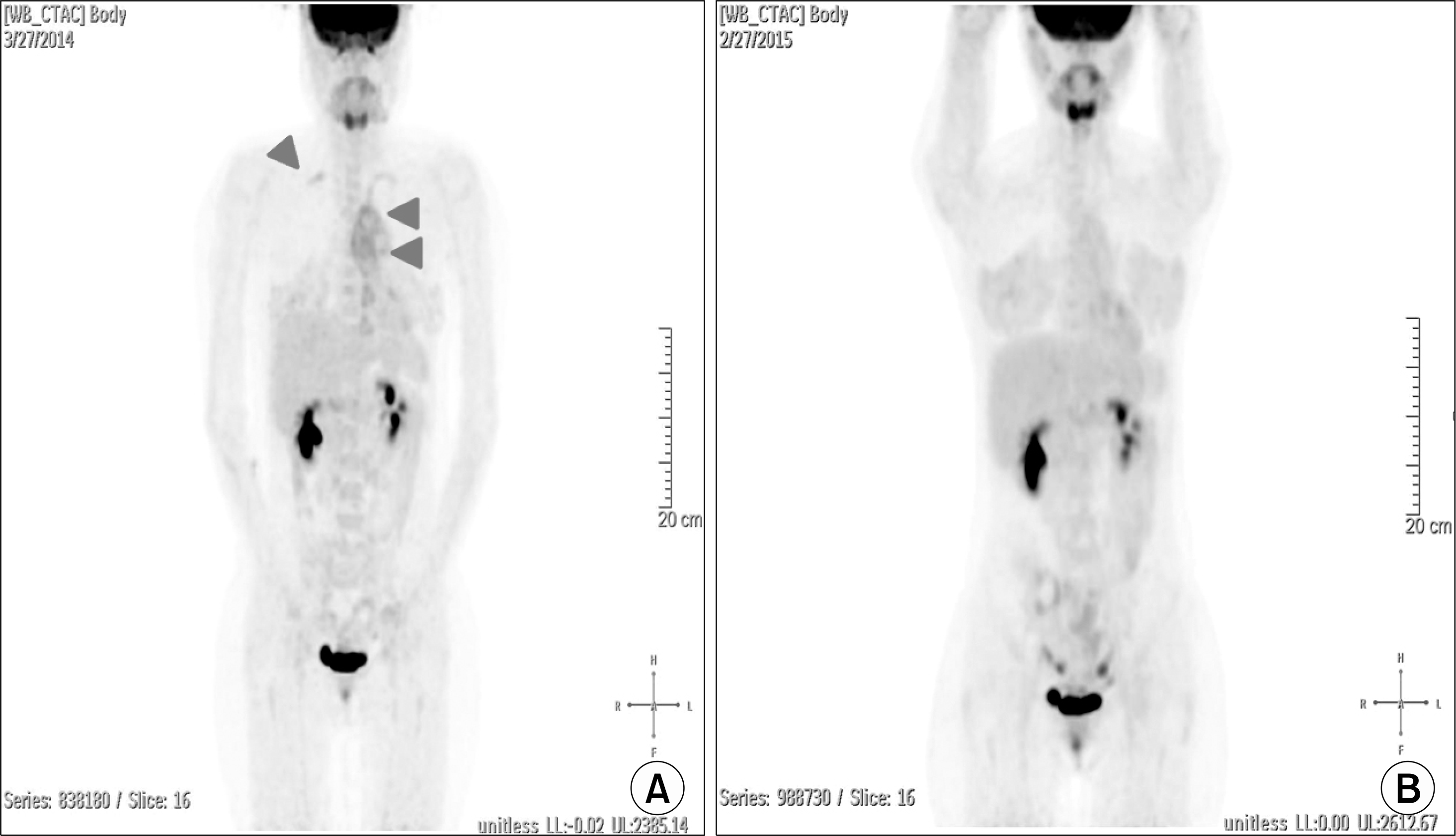 | Figure 3.(A) Before infliximab treatment; 18 F-Fluoro-2-deoxy-D-glucose (18 F-FDG) positron emission tomography scan shows increased uptake of 18 F-FDG in the aortic arch and bilateral subclavian arteritis (arrowheads; visual grade=3, the number of active vascular lesions=5). (B) Twelve months after infliximab treatment: decreased uptake of 18 F-FDG is evident in the inflamed arterial walls (visual grade=1, the number of active vascular lesions=0). |
 XML Download