

 PDF
PDF ePub
ePub Citation
 Print
Print


REFERENCES
1. Yazici H, Fresko I, Yurdakul S. Behçet's syndrome: disease manifestations, management, and advances in treatment. Nat Clin Pract Rheumatol. 2007; 3:148–55.

2. Kim JS. Moyamoya disease: epidemiology, clinical features, and diagnosis. J Stroke. 2016; 18:2–11.
3. Chen JB, Liu Y, Zhou LX, Sun H, He M, You C. Prevalence of autoimmune disease in moyamoya disease patients in Western Chinese population. J Neurol Sci. 2015; 351:184–6.
4. Park YW, Chung JW, Yoon HJ, Yoon W, Kee SJ, Lee SS. Behçet's disease presenting with clinical manifestations of moyamoya disease. J Korean Rheum Assoc. 2005; 12:227–30.
5. Joo SP, Kim TS, Lee JH, Lee JK, Kim JH, Kim SH, et al. Moyamoya disease associated with Behcet's disease. J Clin Neurosci. 2006; 13:364–7.
6. Degirmenci E, Bir LS, Yagcı B, Nazliel B, Siva A. Moyamoya syndrome or Behçet's disease? Int J Rheum Dis. 2014; 17:920–2.
7. Fei Y, Li X, Lin S, Song X, Wu Q, Zhu Y, et al. Major vascular involvement in Behçet's disease: a retrospective study of 796 patients. Clin Rheumatol. 2013; 32:845–52.
8. Han H, Pyo CW, Yoo DS, Huh PW, Cho KS, Kim DS. Associations of Moyamoya patients with HLA class I and class II alleles in the Korean population. J Korean Med Sci. 2003; 18:876–80.
9. Inoue TK, Ikezaki K, Sasazuki T, Matsushima T, Fukui M. Analysis of class II genes of human leukocyte antigen in patients with moyamoya disease. Clin Neurol Neurosurg. 1997; 99(Suppl 2):S234–7.
10. Takeuchi M, Kastner DL, Remmers EF. The immunogenetics of Behçet's disease: A comprehensive review. J Autoimmun. 2015; 64:137–48.
11. Yamada S, Oki K, Itoh Y, Kuroda S, Houkin K, Tominaga T, et al. Effects of surgery and antiplatelet therapy in ten-year follow-up from the registry study of research committee on Moyamoya disease in Japan. J Stroke Cerebrovasc Dis. 2016; 25:340–9.
12. Wang R, Xu Y, Lv R, Chen J. Systemic lupus erythematosus associated with Moyamoya syndrome: a case report and literature review. Lupus. 2013; 22:629–33.
Figure 1.
(A) Initial brain magnetic resonance angiography shows stenosis of bilateral distal internal carotid arteries with well-developed basal and pial collaterals. (B) Initial left internal carotid angiography shows stenosis of the left distal internal carotid artery. Well-developed basal collaterals and pial collaterals to the left hemisphere through the left posterior cerebral artery are also seen. (C) Follow up brain computed tomography shows a newly appearing acute intracerebral hemorrhage in the left thalamus with extension to the ventricles (arrow). (D) On follow up cerebral angiography, aggravated stenosis of the left distal internal carotid artery is seen.

Figure 2.
(A) Abdominal computed tomography shows mild wall thickening of the terminal ileum (arrow) suggesting terminal ileitis. (B) Colonoscopy shows a large ulcerative lesion at the terminal ileum (arrow).
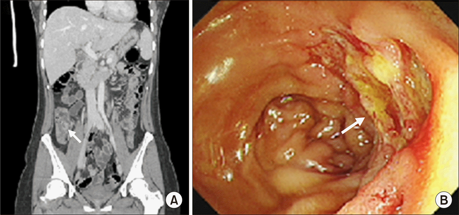
Figure 3.
Acute phase reactant level and medication according to the clinical course. ESR: erythrocyte sedimentation rate, CRP: C-reactive protein, MMD: Moyamoya disease, BD: Behçet's disease, Dx: diagnosed.
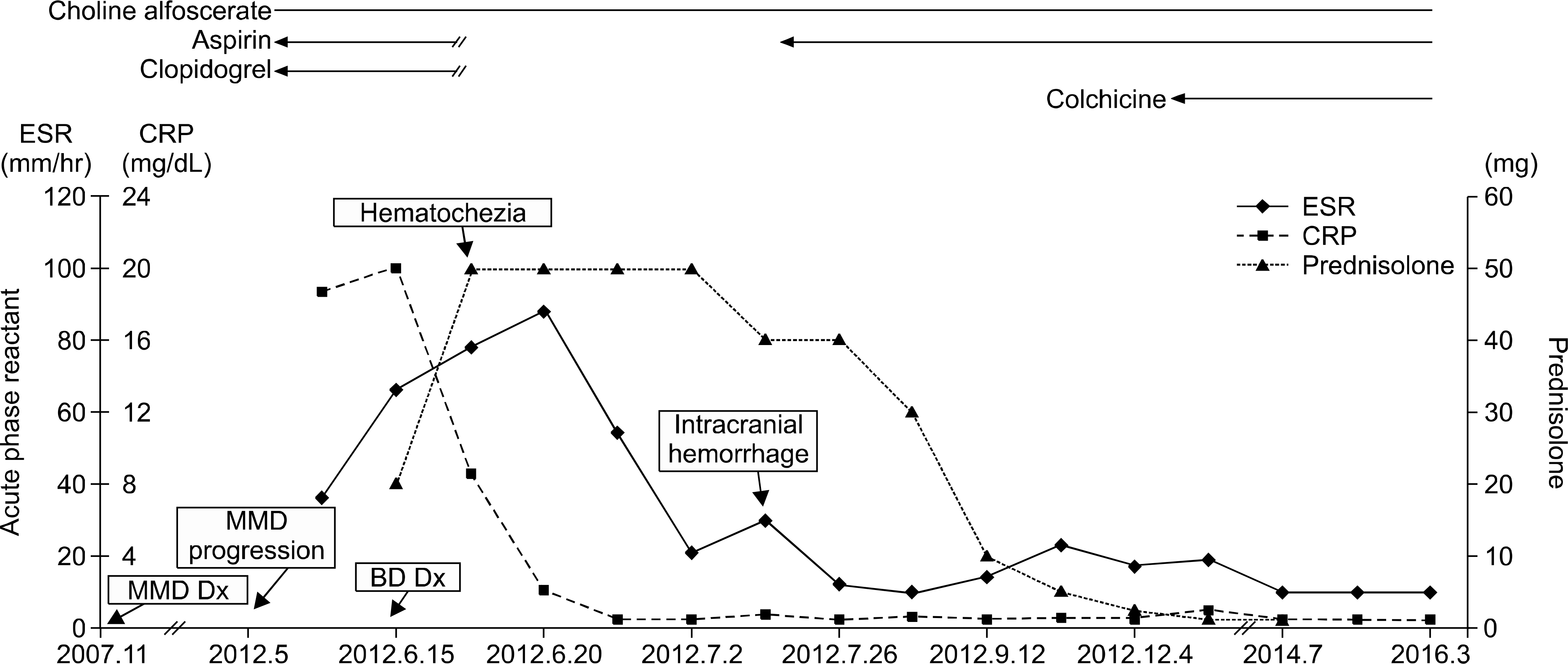
Table 1.
Clinical characteristics of Moyamoya disease (MMD)/Behçet's disease (BD) coexistence cases
 XML Download